Annular Tear
Summary
- Disruption of the annulus fibrosus of an intervertebral disc ("annular fissure" is the preferred nomenclature)
- Usually degenerative, occasionally traumatic
- The hallmark is a T2 high-intensity zone (HIZ) in the posterior annulus, which may enhance
Pathophysiology
- Fissures are classified as concentric (between lamellae), radial (nucleus to outer annulus) or transverse (peripheral, near the ring apophysis)
- Radial fissures are the type that permits disc herniation and are implicated in discogenic pain
Demographics
- Most common in adults aged 30-50 years
- Higher prevalence in:
- Males
- Individuals with physically demanding occupations
- Those with a history of trauma or repetitive stress
Diagnosis
- Clinical presentation:
- Low back pain, often with radicular symptoms
- Pain may worsen with certain movements or positions
- Physical examination:
- Limited range of motion
- Positive straight leg raise test (for lower lumbar tears)
- Diagnostic tests:
- MRI is the gold standard for diagnosis
- Discography may be used in select cases
Imaging
- MRI findings:
- High-intensity zone (HIZ) on T2-weighted images1
- Focal hyperintensity in the posterior annulus on T2-weighted images
- Contrast enhancement of the outer annulus on post-gadolinium T1-weighted images
- CT discography:
- Contrast leakage into the annular tear
- Useful for correlating pain with specific disc levels
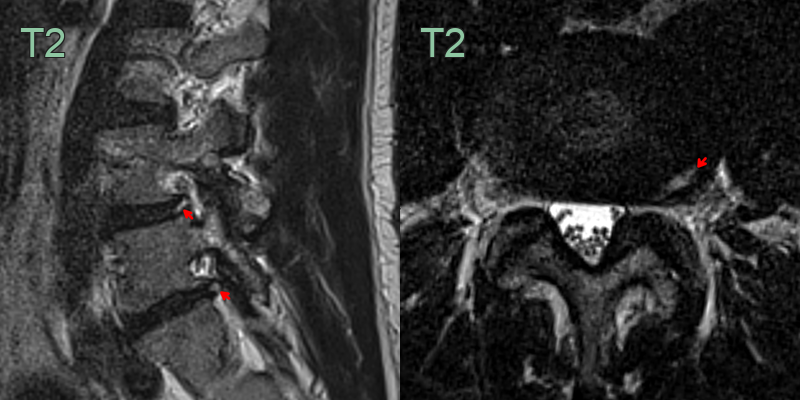
- A 45-year-old patient presented with an acute radiculopathy in the left L4 distribution.
- While there was only minimal intervertebral foraminal narrowing, the L4-5 disc that contained a T2-hyperintense annular tear made contact with the left L4 nerve root.
Treatment
- Conservative: analgesia, activity modification and physiotherapy
- Epidural steroid injection or surgery reserved for refractory radiculopathy
Differential diagnosis
| Imaging differential | Differentiating feature |
|---|---|
| Disc herniation | Focal protrusion/extrusion of disc material beyond the annulus, often with nerve root contact |
| Modic type 1 endplate change | Endplate T1 hypointensity/T2 hyperintensity, distinct from an intra-annular HIZ |
| Discitis/osteomyelitis | Endplate erosion, disc-space T2 hyperintensity and rim-enhancing collection |
| Vertebral metastasis | Enhancing marrow-replacing lesion with cortical destruction |
-
Hadjipavlou et al. Pathomechanics and clinical relevance of disc degeneration and annular tear: a point-of-view review. 1999. American journal of orthopedics (Belle Mead, N.J.) - Open in new tab. ↩