Benign Parietal Thinning
Summary
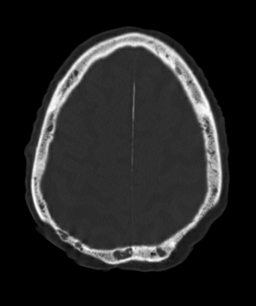
- Benign parietal thinning is a focal thinning of the parietal bone, typically bilateral and symmetrical
- Asymptomatic condition, often discovered incidentally on imaging studies
- No intervention required; important to differentiate from pathological conditions
Pathophysiology
- Exact etiology remains unclear
- Hypothesized mechanisms:
- Congenital developmental variation
- Age-related bone resorption
- Hormonal influences, particularly in postmenopausal women
- Characterised by focal thinning of the outer table and diploe, with preservation of the inner table
Demographics
- More common in older adults, typically over 60 years of age
- Higher prevalence in females, particularly postmenopausal women
- No known racial or ethnic predisposition
- Estimated prevalence of 0.4-1.3% in the general population
Diagnosis
- Usually an incidental finding on imaging studies
- Clinical presentation:
- Asymptomatic
- No associated neurological deficits or palpable abnormalities
- Differential diagnosis:
- Metastatic lesions
- Multiple myeloma
- Hyperparathyroidism
- Fibrous dysplasia
- Paget's disease
Imaging
- CT:
- Bilateral, symmetrical focal thinning of the parietal bones
- Typically located in the parasagittal region
- Sharply demarcated margins
- Preservation of the inner table
- No associated soft tissue mass or bone destruction
- MRI:
- T1 and T2 signal intensity similar to normal bone marrow
- No contrast enhancement
- Skull radiographs:
- May show focal lucencies in the parietal region
- Less sensitive than CT for detection and characterization
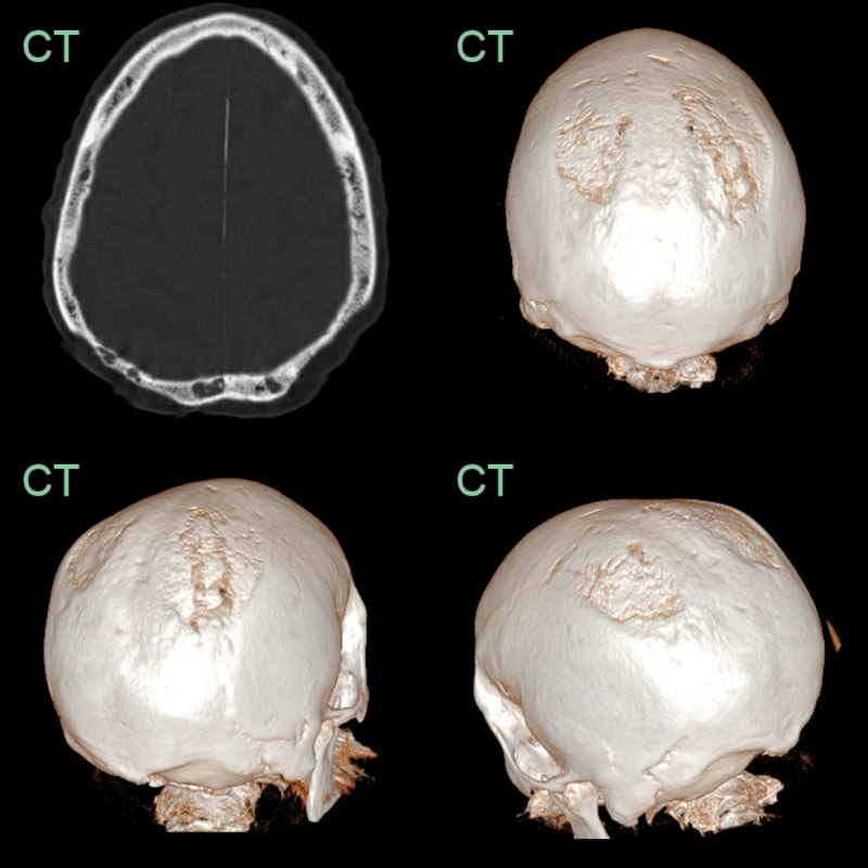
- Incidental finding of thinning of the parietal bone with loss of the outer cortex that was stable over at least 5 years.
- The patient was systemically well with no clinical or laboratory evidence of a haematological disorder.
Treatment
- None required; recognition avoids mistaking it for a destructive lesion1
Differential diagnosis
| Imaging differential | Differentiating feature |
|---|---|
| Lytic metastasis / myeloma | Asymmetric, permeative or "punched-out" lucencies with a soft-tissue component and inner-table destruction |
| Parietal skull fracture | Sharp linear lucency without smooth thinning; overlying scalp swelling |
| Parietal foramina | Well-corticated defects, usually paired and more posterior |
-
Sanati-Mehrizy et al. Characterization of Bilateral Parietal Thinning. 2020. The Journal of craniofacial surgery - Open in new tab. ↩