CSF-venous fistula
Summary
- Abnormal communication between CSF space and venous system
- Causes spontaneous intracranial hypotension (SIH)
- Diagnosed by specialised imaging techniques, often missed on routine studies
Pathophysiology
- Direct connection between CSF and venous compartments
- Often at nerve root sleeve level
- Can occur at spinal or skull base level
- Results in CSF leakage and intracranial hypotension
- Proposed mechanisms:
- Congenital weakness in dura mater
- Trauma or iatrogenic causes
- Degenerative changes in spinal structures
Demographics
- More common in middle-aged adults (40-60 years)
- Slight female predominance
- Associated conditions:
- Connective tissue disorders (e.g., Ehlers-Danlos syndrome)
- History of spinal surgery or intervention
Diagnosis
- Clinical presentation:
- Orthostatic headache
- Neck pain or stiffness
- Tinnitus
- Visual disturbances
- Cerebrospinal fluid analysis:
- Often normal or shows mildly low opening pressure
- May have slightly elevated protein levels
- High clinical suspicion required due to subtle nature of fistulas1
Imaging
- Conventional MRI:
- Brain: pachymeningeal enhancement, subdural collections, pituitary enlargement
- Spine: may show extradural fluid collections
- CT myelography:
- Limited sensitivity for small fistulas
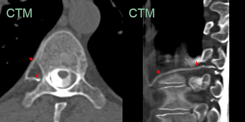
- Lateral decubitus digital subtraction myelography or dynamic CT myelography is the key test, imaging the dependent side
- The "hyperdense paraspinal vein sign" (early opacification of a paraspinal vein) localises the fistula
- Routine spine MRI is typically normal (no extradural fluid), which distinguishes a fistula from a dural CSF leak
Treatment
- Unlike a dural leak, a CSF-venous fistula responds poorly to blood patching
- Definitive treatment is transvenous embolisation or surgical ligation of the draining vein
Differential diagnosis
| Differential Diagnosis | Differentiating Feature |
|---|---|
| CSF leak | Epidural CSF collection often associated with an osteophyte |
| Chiari malformation | Cerebellar tonsillar herniation on MRI |
-
Roytman et al. CSF-Venous Fistula. 2021. Current pain and headache reports - Open in new tab. ↩