Ischaemic Stroke
Summary
- Acute neurological deficit due to focal brain ischaemia
- Caused by thrombotic or embolic occlusion of cerebral arteries
- Imaging crucial for diagnosis, treatment planning, and prognosis
Pathophysiology
- Interruption of blood supply to brain tissue
- Thrombotic: In-situ clot formation due to atherosclerosis
- Embolic: Clot travels from another site (e.g., heart, carotid arteries)
- Ischaemic cascade leads to neuronal death
- Energy failure, excitotoxicity, oxidative stress, inflammation
- Penumbra: potentially salvageable tissue surrounding infarct core
Demographics
- Incidence increases with age
- Risk factors :
- Hypertension
- Diabetes mellitus
- Smoking
- Atrial fibrillation
- Hyperlipidaemia
- Obesity
- Higher prevalence in men, but more severe in women
Diagnosis
- Clinical presentation: sudden onset of focal neurological deficits
- National Institutes of Health Stroke Scale (NIHSS) for severity assessment
- Laboratory tests: complete blood count, coagulation profile, lipid panel
- Electrocardiogram to detect atrial fibrillation
- Imaging essential for definitive diagnosis and treatment planning
Imaging
- Non-contrast CT :
- First-line imaging modality
- Excludes haemorrhage
- Early signs: hyperdense vessel sign, loss of gray-white matter differentiation
- CT angiography:
- Identifies site of vessel occlusion
- Evaluates collateral circulation
- CT perfusion:
- Assesses penumbra and infarct core
- Guides thrombectomy decision-making
- MRI :
- Higher sensitivity for acute infarction
- Diffusion-weighted imaging (DWI): early detection of cytotoxic oedema
- FLAIR: subacute to chronic infarcts
- Susceptibility-weighted imaging: microbleeds, thrombus
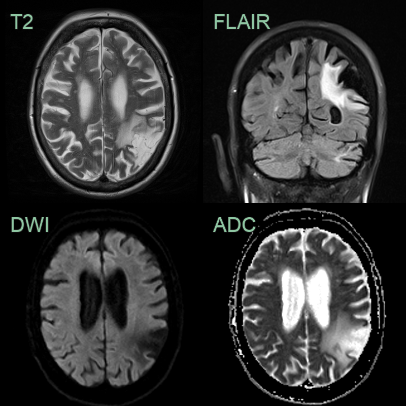
- Mature infarct in the left MCA territory with facilitated diffusion, gliotic T2-hyperintensity, and volume loss.
Treatment
- IV thrombolysis within 4.5 hours and mechanical thrombectomy for large-vessel occlusion (up to 24 hours with imaging selection); then secondary prevention1
Differential diagnosis
| Differential Diagnosis | Distinguishing Feature |
|---|---|
| Encephalitis | May cross arterial territories, ADC values may not be so low, fever/altered mental status/'active' CSF |
-
Tsivgoulis et al. Thrombolysis for acute ischaemic stroke: current status and future perspectives. 2023. The Lancet. Neurology - Open in new tab. ↩