Kikuchi-Fujimoto Disease
Summary
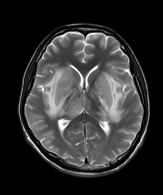
- Rare, self-limiting condition characterised by cervical lymphadenopathy and fever
- Benign histiocytic necrotising lymphadenitis with unknown aetiology
- Diagnosis based on clinical presentation, lymph node biopsy, and exclusion of other causes
Pathophysiology
- Exact cause unknown, but likely an autoimmune response to an infectious trigger
- Proposed mechanisms:
- Viral infection (e.g., Epstein-Barr virus, human herpesvirus 6)
- Autoimmune disorder (association with systemic lupus erythematosus)
- Histopathology shows:
- Paracortical necrosis with karyorrhectic debris
- Histiocytic infiltrate with absence of neutrophils
- Proliferation of CD68+ histiocytes and CD8+ T-lymphocytes
Demographics
- Predominantly affects young adults (20-30 years old)
- Female preponderance (female to male ratio 4:1)
- More common in Asian populations, but can occur in any ethnic group
- Rare in children and elderly
Diagnosis
- Clinical presentation:
- Cervical lymphadenopathy (unilateral or bilateral)1
- Fever
- Night sweats
- Fatigue
- Weight loss
- Laboratory findings:
- Leukopenia
- Elevated erythrocyte sedimentation rate (ESR)
- Elevated C-reactive protein (CRP)
- Definitive diagnosis:
- Excisional lymph node biopsy with characteristic histopathological findings
- Differential diagnosis:
- Lymphoma
- Tuberculosis
- Systemic lupus erythematosus
- Cat scratch disease
- Toxoplasmosis
Imaging
- Ultrasonography:
- Enlarged hypoechoic lymph nodes
- Preservation of hilar vascularity
- Cortical thickening
- Computed Tomography (CT):
- Multiple enlarged lymph nodes, typically in cervical region
- Homogeneous enhancement
- Absence of necrosis or extracapsular spread
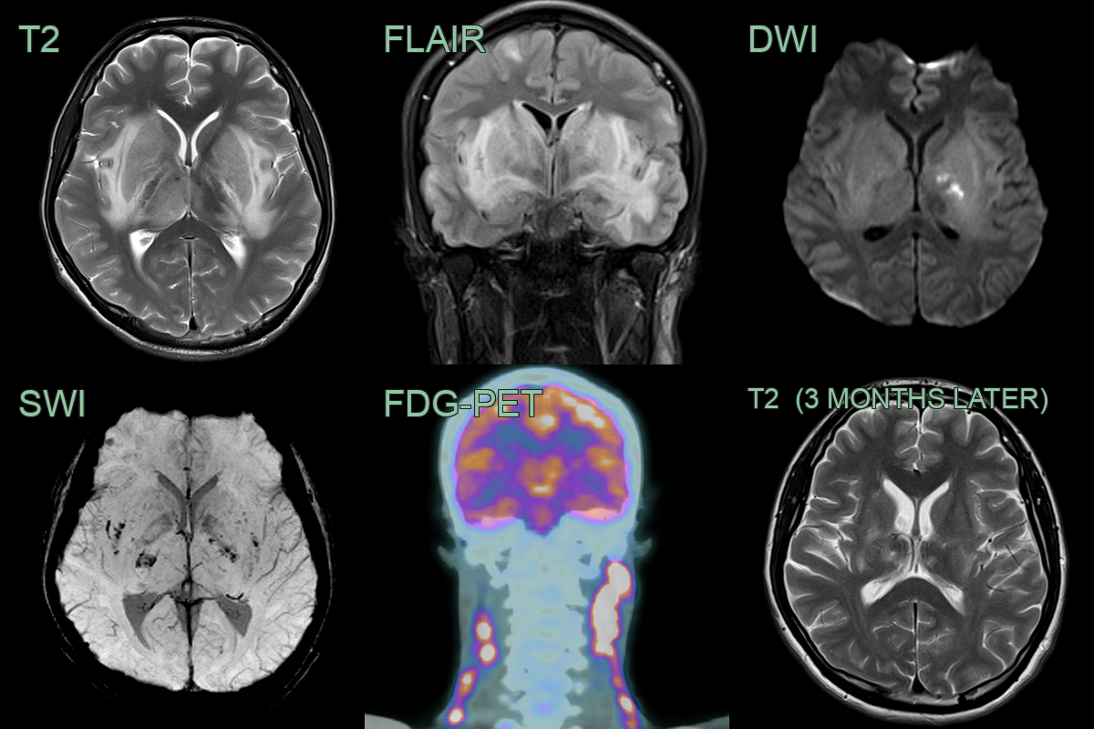
- Magnetic Resonance Imaging (MRI):
- T1: Isointense to muscle
- T2: Hyperintense
- Post-contrast: Homogeneous enhancement
- Diffusion-weighted imaging: Restricted diffusion
- 18F-FDG PET/CT:
- Increased FDG uptake in affected lymph nodes
- Useful for excluding malignancy and monitoring treatment response
Treatment
- Self-limiting; supportive care, with steroids for severe cases. Follow-up is warranted as it can herald SLE
Differential diagnosis (cervical lymphadenopathy)
| Imaging differential | Differentiating feature |
|---|---|
| Lymphoma | Bulky, often bilateral nodes; KFD nodes are usually smaller and unilateral (posterior triangle) with preserved architecture |
| Tuberculous adenitis | Central necrosis and rim enhancement; conglomerate nodes |
| Metastatic nodes | Necrosis, irregular margins and a known primary; extracapsular spread |
| Reactive/suppurative adenitis | Preserved hilum; frank abscess if suppurative |
-
Perry et al. Kikuchi-Fujimoto Disease: A Review. 2018. Archives of pathology & laboratory medicine - Open in new tab. ↩