Leptomeningeal Carcinomatosis
Summary
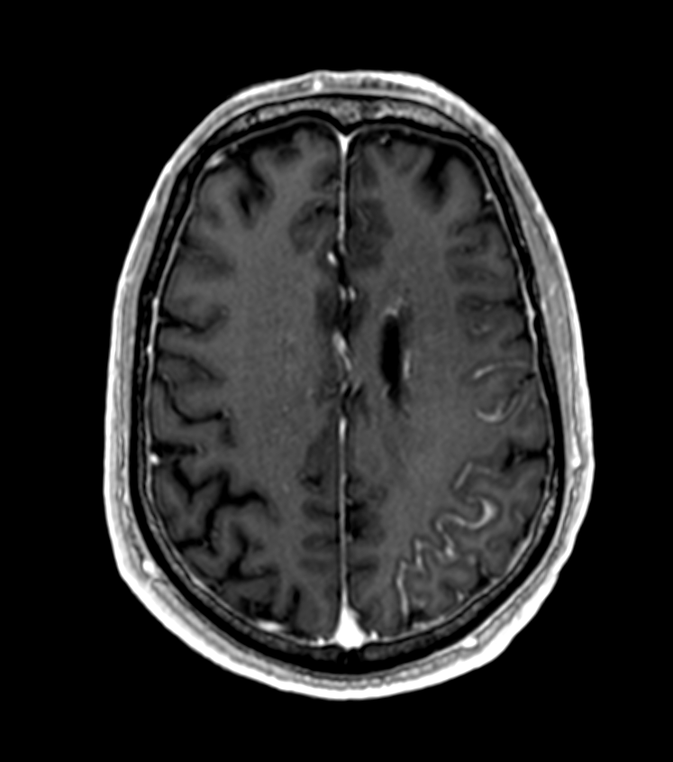
- Leptomeningeal carcinomatosis (LC) is the spread of malignant cells to the leptomeninges and subarachnoid space
- Presents with multifocal neurological symptoms and signs
- Diagnosis relies on CSF cytology and neuroimaging, particularly contrast-enhanced MRI
Pathophysiology
- Malignant cells reach the leptomeninges via:
- Haematogenous spread
- Direct extension from brain or spinal cord metastases
- Perineural or perivascular spread
- Tumour cells proliferate in the subarachnoid space, leading to:
- Obstruction of CSF flow
- Infiltration of cranial and spinal nerve roots
- Invasion of brain parenchyma
Demographics
- Occurs in 5-8% of patients with solid tumours
- Most common primary tumours:
- Breast cancer (12-35%)
- Lung cancer (10-26%)
- Melanoma (5-25%)
- Incidence increasing due to improved survival of cancer patients and better diagnostic techniques
Diagnosis
- Clinical presentation:
- Headache
- Altered mental status
- Cranial nerve palsies
- Radicular pain
- Cauda equina syndrome
- CSF analysis:
- Cytology (gold standard)
- Elevated protein
- Decreased glucose
- Increased opening pressure
- Neuroimaging (MRI with gadolinium)1
- Meningeal biopsy (rarely required)
Imaging
- MRI with gadolinium is the imaging modality of choice
- Findings:
- Leptomeningeal enhancement
- Nodular or linear enhancement along the surface of the brain and spinal cord
- Hydrocephalus
- Subarachnoid nodules
- Cranial nerve enhancement
- CT with contrast:
- Less sensitive than MRI
- May show leptomeningeal enhancement or hydrocephalus
- FDG-PET:
- Can detect metabolically active leptomeningeal disease
- Limited sensitivity for small volume disease
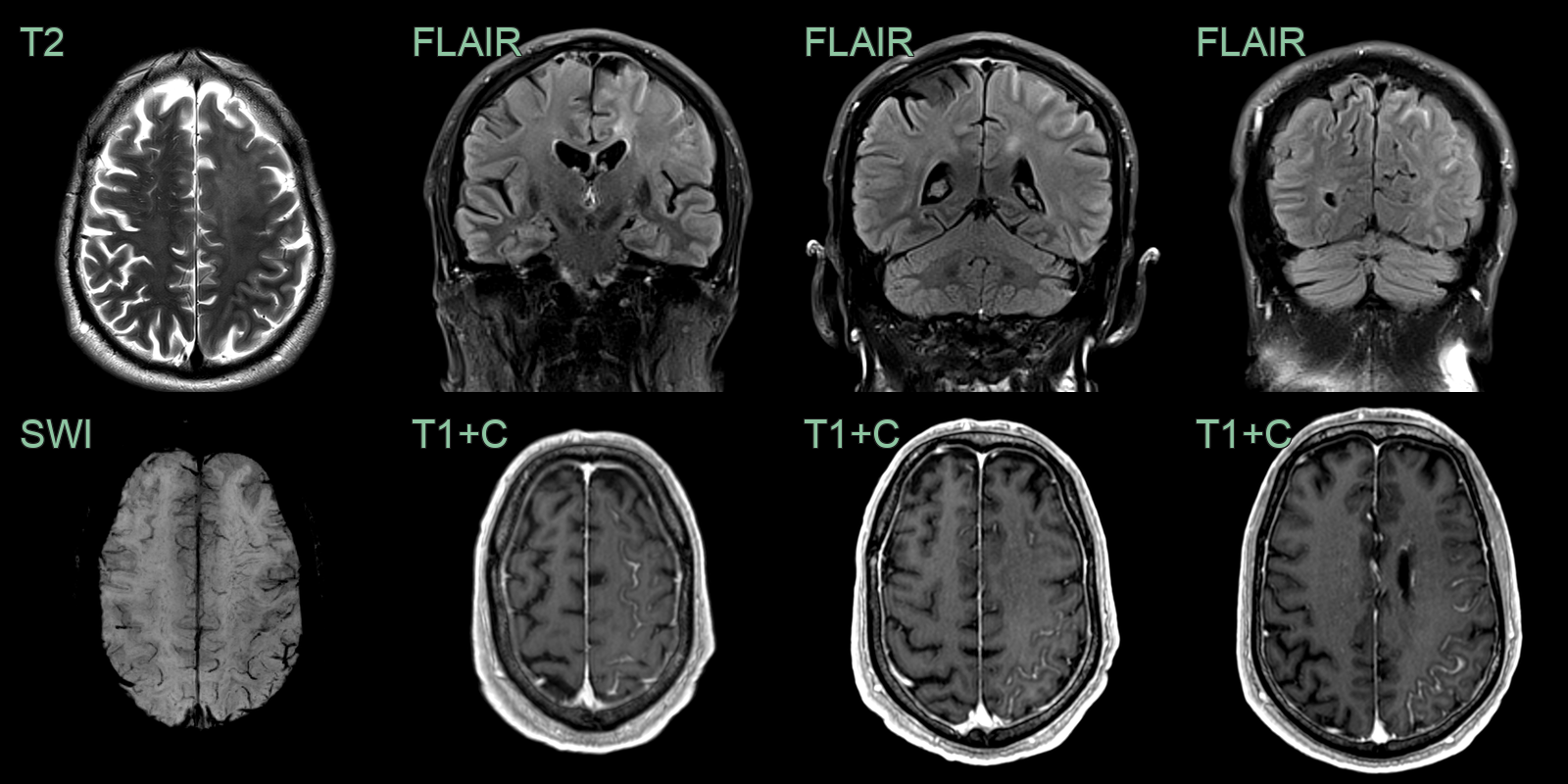
- A 70-year-old patient with metastatic prostate cancer presented with right arm weakness.
- MRI showed hazy T2-hyperintensity within the right frontal lobe and evidence of venous congestion on SWI.
- Sulcal FLAIR hyperintensity and enhancement indicated leptomeningeal carcinomatosis.
Treatment
- Intrathecal/systemic chemotherapy and radiotherapy to symptomatic sites; prognosis is poor. Whole-neuraxis MRI plus CSF cytology is the diagnostic combination
Differential diagnosis (leptomeningeal enhancement)
| Imaging differential | Differentiating feature |
|---|---|
| Infectious (bacterial) meningitis | Diffuse smooth pial enhancement with clinical sepsis and active CSF |
| Tuberculous meningitis | Thick basal enhancement with perforator infarcts and hydrocephalus |
| Neurosarcoidosis | Nodular pial and dural enhancement, often basal, with hypothalamic/stalk involvement |
| Reactive/post-procedural | Thin smooth enhancement after lumbar puncture or shunting, without nodules |
-
Grossman et al. Leptomeningeal carcinomatosis. 1999. Cancer treatment reviews - Open in new tab. ↩