Pineal Tumour of Intermediate Differentiation
Summary
- Rare neoplasm of the pineal gland with mixed features of pineocytoma and pineoblastoma
- Clinical presentation includes headache, visual disturbances, and Parinaud syndrome
- Imaging shows a well-defined, heterogeneous mass in the pineal region with variable enhancement
Pathophysiology
- Arises from pineal parenchymal cells
- WHO grade 2 or 3 tumour, depending on mitotic activity and neurofilament protein expression
- Exhibits intermediate differentiation between pineocytoma and pineoblastoma
- May show areas of necrosis and calcification
Demographics
- Rare tumour, accounting for <0.1% of all intracranial neoplasms
- Typically affects young adults and middle-aged individuals
- No significant gender predilection reported
Diagnosis
- Clinical presentation:
- Headache
- Visual disturbances
- Parinaud syndrome (vertical gaze palsy, convergence-retraction nystagmus)
- Hydrocephalus due to compression of the cerebral aqueduct
- Histopathology:
- Moderate cellularity with diffuse growth pattern
- Intermediate nuclear-to-cytoplasmic ratio
- Mild to moderate nuclear atypia
- Immunohistochemistry positive for synaptophysin and neurofilament protein
Imaging
- CT:
- Well-defined, hyperdense mass in the pineal region1
- Variable calcification patterns
- Contrast enhancement may be heterogeneous
- MRI:
- T1: Iso- to hypointense
- T2: Iso- to hyperintense
- T1 post-contrast: Heterogeneous enhancement
- DWI: Variable restricted diffusion
- MR spectroscopy: Elevated choline peak, reduced N-acetylaspartate
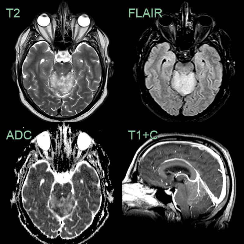
- A 50-year-old patient presented with a 6 month history of occipital headaches.
- MRI showed a lesion in the pineal region. A large vein within the lesion indicated that the lesion was extra-axial.
- Following resection, a pineal tumour of intermediate differentiation (grade 3) was diagnosed.
Treatment
- Resection with adjuvant radiotherapy; craniospinal irradiation for disseminated disease
Differential diagnosis (pineal region mass)
| Imaging differential | Differentiating feature |
|---|---|
| Pineoblastoma | Larger, more heterogeneous and aggressive, with "exploded" peripheral calcification and marked diffusion restriction (WHO grade 4) |
| Pineocytoma | Small (<3 cm), well-defined, often cystic, indolent (WHO grade 1) |
| Germinoma | Engulfed central calcification, avid homogeneous enhancement and restricted diffusion, in a young male, with raised markers (β-hCG/PLAP) |
| Pineal cyst | Thin-walled CSF-signal cyst without a solid enhancing nodule |
| Tectal/pineal region glioma | Arises from the tectum, expanding it, rather than a discrete pineal mass |
-
Takase et al. Pineal parenchymal tumor of intermediate differentiation: a systematic review and contemporary management of 389 cases reported during the last two decades. 2022. Neurosurgical review - Open in new tab. ↩