Vertebral Artery Dissection
Summary
- Vertebral artery dissection (VAD) is a tear in the inner lining of the vertebral artery, leading to intramural haematoma formation
- Presents with neck pain, headache, and posterior circulation ischaemic symptoms
- Diagnosis relies on clinical presentation and neuroimaging findings
Pathophysiology
- Intimal tear allows blood to enter the arterial wall, creating a false lumen
- Mechanisms:
- Spontaneous: often associated with connective tissue disorders
- Traumatic: minor trauma or sudden neck movements
- Consequences:
- Luminal stenosis or occlusion
- Thrombus formation and distal embolisation
- Subarachnoid haemorrhage (rare)
Demographics
- Incidence: 1-1.5 per 100,000 per year
- Age: typically affects young to middle-aged adults (mean age 40-45 years)
- Gender: slight female predominance
- Risk factors:
- Hypertension
- Smoking
- Migraine
- Oral contraceptive use
- Connective tissue disorders (e.g., Ehlers-Danlos syndrome, Marfan syndrome)
Diagnosis
- Clinical presentation:
- Neck pain or occipital headache (often unilateral)1
- Posterior circulation ischaemic symptoms:
- Vertigo
- Diplopia
- Ataxia
- Lateral medullary syndrome (Wallenberg syndrome)
- Physical examination:
- Horner's syndrome (ipsilateral miosis, ptosis, and anhidrosis)
- Nystagmus
- Cerebellar signs
- Laboratory tests:
- No specific blood markers for VAD
- Consider testing for connective tissue disorders if suspected
Imaging
- CT angiography (CTA):
- First-line imaging modality
- Findings:
- Eccentric luminal narrowing
- Intramural haematoma (crescent sign)
- Vessel irregularity or pseudoaneurysm formation
- MRI and MR angiography (MRA):
- Superior soft tissue contrast
- Findings:
- Intramural haematoma: hyperintense on T1-weighted fat-suppressed images
- Luminal narrowing or occlusion on MRA
- Digital subtraction angiography (DSA):
- Gold standard for diagnosis
- Reserved for cases with equivocal non-invasive imaging or when endovascular treatment is planned
- Findings:
- String sign (long, irregular narrowing)
- Pearl-and-string sign (alternating dilatations and stenoses)
- Pseudoaneurysm formation
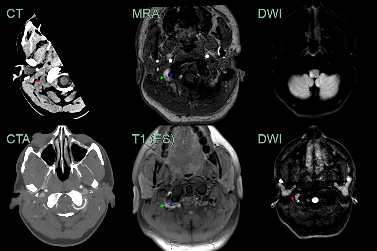
- A 30-year-old patient presented with dizziness, nystagmus, dysphagia and ataxia.
- CT showed a subtly hyperdense rim around the left V3 vertebral artery (red arrow).
- The corresponding thrombus caused DWI hyperintensity (red arrow) and T1-shortening on the ToF angiogram (red arrow) next to flow-related signal from the narrowed lumen (blue arrow).
- The Wallenberg syndrome was caused by a lateral medullary infarct.
Treatment
- Antiplatelet or anticoagulation to prevent thromboembolic (posterior circulation) stroke; endovascular treatment for refractory cases
- The crescentic T1-hyperintense intramural haematoma on fat-saturated imaging is the most specific sign
-
Clark et al. A review of carotid and vertebral artery dissection. 2022. British journal of hospital medicine (London, England : 2005) - Open in new tab. ↩