Cross Cerebellar Diaschisis
Summary
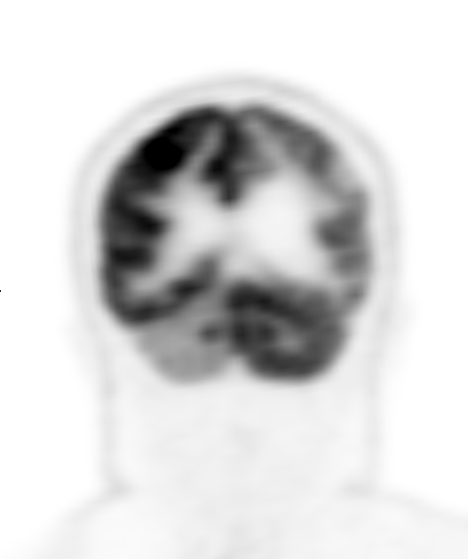
- Functional depression of the contralateral cerebellar hemisphere following a supratentorial lesion
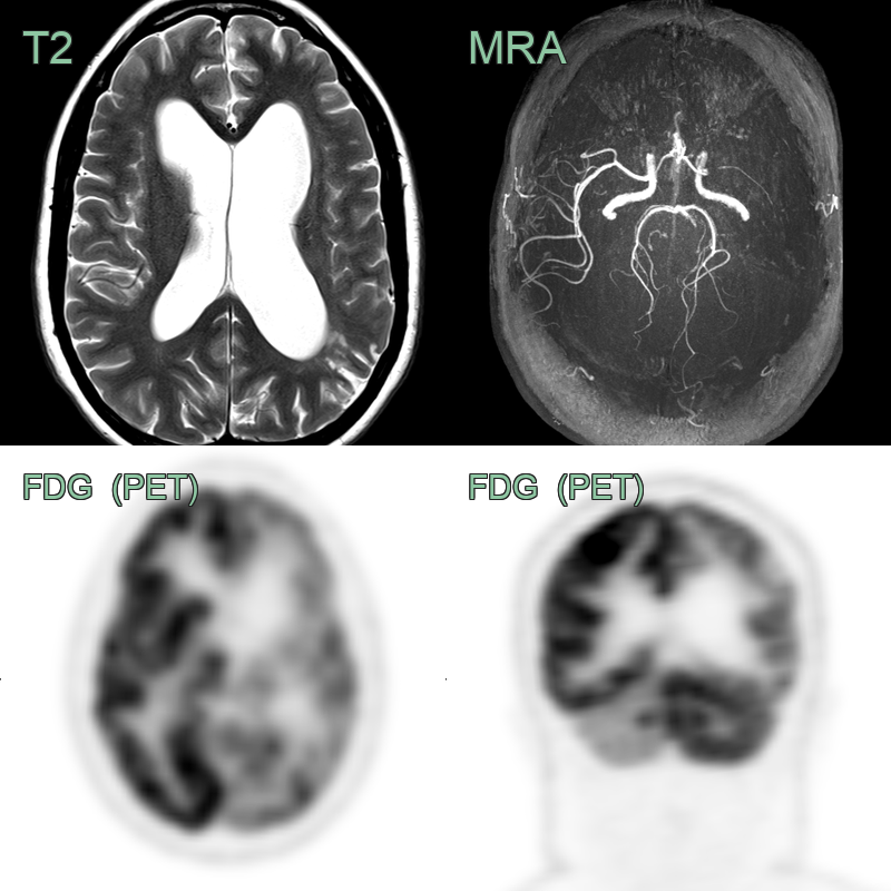
- Characterised by reduced blood flow and metabolism in the cerebellar hemisphere opposite to a focal supratentorial lesion
- Typically associated with stroke but can occur in other conditions affecting cerebral cortex or subcortical structures
Mechanism
- Disruption of the corticopontocerebellar pathway leads to deafferentation of the contralateral cerebellar hemisphere1
- Reduced excitatory input from the cerebral cortex results in decreased neuronal activity, blood flow and metabolism in the affected cerebellar hemisphere
- Mediated by transneuronal metabolic depression rather than direct ischaemia
Imaging Appearance
- Conventional CT and MRI:
- Usually normal in the affected cerebellar hemisphere
- Useful for identifying the primary supratentorial lesion
- SPECT (Single Photon Emission Computed Tomography):
- Gold standard for diagnosis
- Shows decreased perfusion in the contralateral cerebellar hemisphere
- PET (Positron Emission Tomography):
- Demonstrates reduced glucose metabolism in the affected cerebellar hemisphere
- CT perfusion:
- May show decreased blood flow in the contralateral cerebellar hemisphere
- MRI:
- Arterial spin labeling (ASL) perfusion can demonstrate reduced cerebellar blood flow
- Diffusion-weighted imaging (DWI) may show restricted diffusion in acute cases
Clinical Relevance
- Not a lesion in its own right and requires no specific treatment: recognising it avoids misinterpreting the cerebellar asymmetry as a separate lesion
- Most commonly seen with acute ischaemic stroke, particularly in the middle cerebral artery territory, and with large hemispheric lesions or subcortical white matter and basal ganglia involvement
- Also observed in traumatic brain injury, tumours and epilepsy
- Usually clinically occult, as symptoms are masked by the primary supratentorial lesion; recognition therefore relies on functional neuroimaging
Differential diagnosis
| Differential Diagnosis | Differentiating Feature |
|---|---|
| Cerebellar infarction | Restricted diffusion on DWI; does not cross midline |
| Posterior reversible encephalopathy syndrome (PRES) | Bilateral involvement, often in parieto-occipital regions |
| Cerebellar tumour | Mass effect, enhancement on contrast-enhanced MRI |
| Cerebellar abscess | Ring-enhancing lesion with restricted diffusion |
| Wernicke encephalopathy | Bilateral symmetrical involvement of mammillary bodies, thalami, and periaqueductal gray matter |
| Cerebellar atrophy | Generalized volume loss, not unilateral |
| Multiple sclerosis | Multiple white matter lesions, often ovoid and periventricular |
| Cerebellar contusion | Associated haemorrhagic foci on GRE/SWI; overlying skull fracture or extracranial soft tissue swelling on CT |
| Metastatic disease | Multiple lesions with surrounding oedema; ring or nodular enhancement; no ipsilateral supratentorial lesion |
| Spinocerebellar ataxia | Bilateral symmetric cerebellar and brainstem atrophy; no corresponding supratentorial lesion |
-
M Reivich. Crossed cerebellar diaschisis. 1992. AJNR. American journal of neuroradiology - Open in new tab. ↩