CTA Spot Sign
Summary
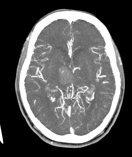
- One or more foci of contrast within an acute intracerebral haematoma on CT angiography, representing active contrast extravasation
- A strong predictor of haematoma expansion and is associated with worse clinical outcome
- Distinct from the CTA "dot sign", which is intraluminal thrombus in a distal MCA branch in acute ischaemic stroke
Imaging Appearance
- Basis of the sign:
- Active extravasation of contrast from ruptured small vessels within an acute intracerebral haematoma, reflecting ongoing bleeding1
- Identified on CT angiography source images, with commonly applied criteria for a spot:
- One or more foci of contrast within the haematoma
- Discontinuous from adjacent normal or abnormal vasculature
- Attenuation of at least 120 HU
- Any size or morphology
- Delayed / post-contrast acquisitions:
- Increase sensitivity by demonstrating contrast leakage or pooling that increases on delayed-phase imaging
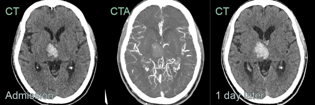
- A 60-year-old patient presented with left sided weakness and paraesthesia.
- Imaging showed a focus of enhancement within a right thalamic haematoma.
- A follow-up CT showed enlargement of the haematoma.
Clinical Relevance
- Most often seen in spontaneous (primary) intracerebral haemorrhage, including hypertensive and cerebral amyloid angiopathy-related bleeds
- More frequent with larger haematomas, earlier imaging after symptom onset, anticoagulant use and coagulopathy
- A prognostic marker rather than a target for direct treatment:
- The number, size and attenuation of spots (the "spot sign score") stratify the risk of expansion and predict haematoma growth on follow-up non-contrast CT
- Identifies patients who warrant close monitoring for haematoma expansion
- Management is directed at the underlying haemorrhage:
- Blood pressure control
- Reversal of anticoagulation and correction of coagulopathy
- Neurosurgical evaluation for evacuation in selected patients
- Note: intravenous thrombolysis is contraindicated in intracerebral haemorrhage
Differential diagnosis
| Imaging differential | Differentiating feature |
|---|---|
| Calcification within the haematoma | Present and dense on the non-contrast scan; does not represent contrast |
| Aneurysm or vascular malformation | Continuous with a vessel or represents an underlying lesion, rather than free extravasation |
| Choroid plexus or vascular calcification | Typical location and present on non-contrast imaging |
| CTA dot sign | Intraluminal thrombus in a distal MCA branch in acute ischaemic stroke, not within a haematoma |
-
Peng et al. Predictive Value of CTA Spot Sign on Hematoma Expansion in Intracerebral Hemorrhage Patients. 2017. BioMed research international - Open in new tab. ↩