Paramagnetic Rim Lesions
Summary
- Paramagnetic rim lesions (PRLs) are chronic active demyelinating lesions characterized by a peripheral rim of iron-laden microglia/macrophages.
- On susceptibility-based MRI, they appear as white matter lesions with a hypointense paramagnetic rim.
- PRLs are an emerging imaging biomarker of chronic active inflammation in multiple sclerosis (MS).
Imaging Appearance
- Susceptibility-weighted imaging (SWI):
- Characteristic peripheral hypointense (paramagnetic) rim surrounding an iso- to hypointense lesion core.
- Rim reflects iron deposition within activated microglia/macrophages at the lesion edge.
- Quantitative susceptibility mapping (QSM):
- Considered the most specific sequence; rim appears hyperintense (paramagnetic signal) with a non-paramagnetic core.
- Helps distinguish iron (paramagnetic) from other sources of signal.
- T2*/gradient-echo phase imaging:
- Rim demonstrates paramagnetic phase shift.
- Field strength:
- Better conspicuity at 7T, but reliably detectable at 3T; 1.5T is less sensitive.
- Distinguishing features:
- Rim is complete or near-complete and persists over time, unlike the transient rim of enhancement seen in acute lesions.
- PRLs are typically non-enhancing on post-contrast T1 (chronic, not acutely inflamed).
- Pitfalls:
- Central vein sign may coexist and supports demyelinating etiology.
- Must differentiate from microbleeds, calcification, and normal deep gray matter iron.
Clinical Relevance
- Marker of chronic active (smoldering) inflammation, associated with ongoing tissue damage and lesion expansion.
- Correlate with greater disability, more aggressive disease course, and worse long-term outcomes in MS.
- Considered highly specific for MS, aiding differentiation from mimics (e.g., migraine, small vessel disease, NMOSD).
- Emerging role as a prognostic and diagnostic biomarker; increasingly incorporated into research and potentially future diagnostic criteria.
- May serve as an imaging endpoint for evaluating therapies targeting compartmentalized/chronic inflammation.
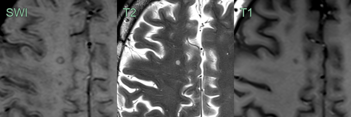
- In the right centrum semiovale, a small well-demarcated T2-hyperintense and T1-hypointense lesion had a rim of susceptibility artefact.