Case of the Month: June 2026
Case history
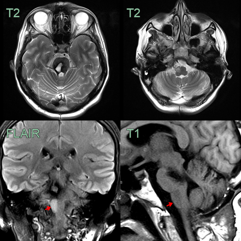
- 45-year-old patient developed a rhythmic palatal tremor one year after resection of a right hemipontine cavernoma.
Click to reveal diagnosis
Hypertrophic Olivary Degeneration
- MRI showed the right inferior olivary nucleus becoming progressively T2-hyperintense and swollen, without diffusion restriction or enhancement.
- Hypertrophic olivary degeneration results from disruption of the dentato-rubro-olivary pathway (the triangle of Guillain-Mollaret), classically presenting with palatal tremor 1-6 months after the inciting lesion.
-
Recognising the hypertrophied, T2-hyperintense olive downstream of a known brainstem or dentate lesion avoids mistaking it for a tumour.