Trigeminal Artery
Summary
- Persistent trigeminal artery (PTA) is the most common persistent carotid-basilar anastomosis
- Embryonic connection between the cavernous portion of the internal carotid artery and the basilar artery
- Usually asymptomatic but can be associated with various neurological symptoms and vascular anomalies
Embryology
- The trigeminal artery develops around the 3rd week of gestation
- It normally regresses by the 7th-8th week of gestation
- A persistent trigeminal artery results when the artery fails to regress and remains patent
Anatomy
- Connects the cavernous segment of the internal carotid artery to the basilar artery
- Two main types:
- Lateral (petrosal) variant: more common; courses lateral to the dorsum sellae
- Medial (sphenoidal) variant: less common; courses medial to the dorsum sellae
- Associated vascular anatomy:
- Hypoplasia of the basilar artery proximal to the PTA junction
- Absence or hypoplasia of the posterior communicating arteries
Imaging
- Angiography (DSA, CTA or MRA):
- Gold standard for diagnosis1
- Demonstrates direct connection between internal carotid artery and basilar artery
- CT:
- May show a rounded or tubular structure in the prepontine cistern
- Calcifications may be present
- MRI:
- T1 and T2-weighted images: Flow void in the prepontine cistern
- MRA: Clearly demonstrates the persistent trigeminal artery
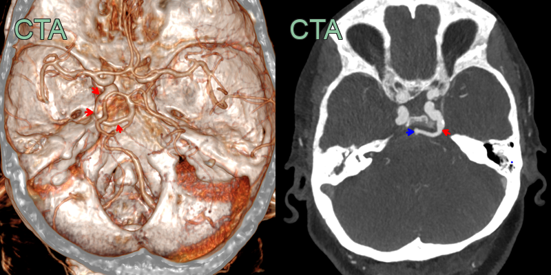
- Incidental finding of a trigeminal artery (red arrow) forming an anastomosis between the cavernous ICA and the basilar artery (blue arrow).
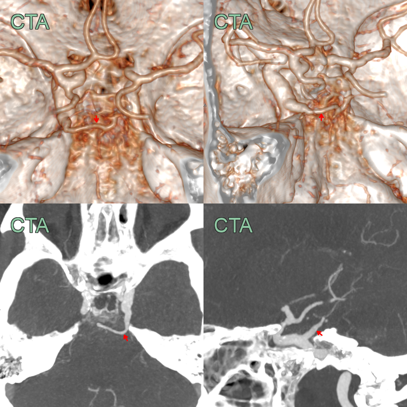
- Incidental finding of an anastomosis between the cavernous left ICA and the posterior circulation (red arrow).
- The vertebrobasilar system was hypoplastic.
Clinical Relevance
- Found in 0.1-0.6% of cerebral angiograms, usually as an incidental finding in adults
- No treatment is required for the variant itself
- Recognition matters because it alters posterior circulation haemodynamics and is associated with aneurysms
- Usually asymptomatic, but reported associations include:
- Trigeminal neuralgia
- Oculomotor palsy
- Abducens nerve palsy
- Cerebrovascular insufficiency
Differential diagnosis
| Differential Diagnosis | Differentiating Feature |
|---|---|
| Persistent Hypoglossal Artery | Arises from cervical internal carotid artery and enters skull through hypoglossal canal |
| Persistent Proatlantal Artery | Connects vertebral artery to external carotid artery |
| Persistent Otic Artery | Passes through internal acoustic meatus |
| Aberrant Internal Carotid Artery | Courses through middle ear cavity |
| Basilar Artery Fenestration | Involves duplication of a segment of the basilar artery |
| Arteriovenous Malformation | Abnormal tangle of blood vessels with direct arterial-venous shunting |
| Aneurysm | Focal dilatation of an artery, typically saccular or fusiform in shape |
| Cavernous Sinus Fistula | Abnormal communication between carotid artery and cavernous sinus |
| Moyamoya Disease | Progressive stenosis of distal internal carotid arteries with collateral vessel formation |
| Basilar Artery Hypoplasia | Underdevelopment of the basilar artery, often associated with persistent fetal circulation |
-
Vasović et al. Trigeminal artery: a review of normal and pathological features. 2012. Child's nervous system : ChNS : official journal of the International Society for Pediatric Neurosurgery - Open in new tab. ↩