Arteriovenous Malformation
Summary
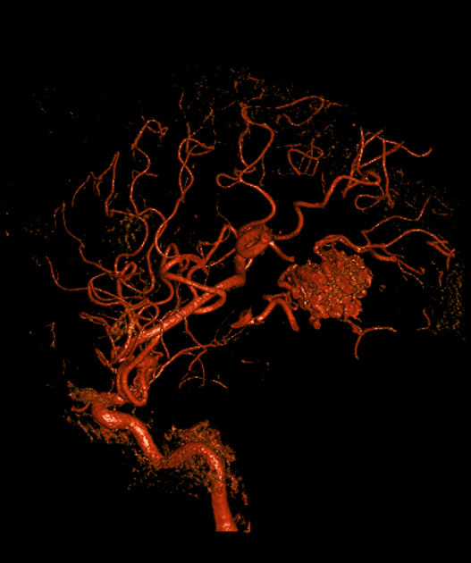
- Congenital vascular anomaly characterised by abnormal connections between arteries and veins, bypassing the capillary bed
- Most commonly occurs in the brain, but can affect any organ system
- Presents with haemorrhage, seizures, or neurological deficits in cerebral AVMs
Pathophysiology
- Abnormal development of embryonic vascular system
- Direct arteriovenous shunting without intervening capillary network
- Progressive dilation of feeding arteries and draining veins
- Risk of rupture due to high-flow, high-pressure system
Demographics
- Prevalence: 18 per 100,000 adults
- Male to female ratio: 1:1
- Most common age of presentation: 20-40 years
- Sporadic occurrence in majority of cases
- Associated with hereditary haemorrhagic telangiectasia in some cases
Diagnosis
- Clinical presentation:
- Intracranial haemorrhage (50%)
- Seizures (30%)
- Headaches (15%)
- Focal neurological deficits (5%)
- Physical examination:
- May be normal
- Focal neurological deficits
- Bruit on auscultation (rare)
- Laboratory tests:
- Generally not specific for AVM diagnosis
Imaging
- Computed Tomography (CT):
- Non-contrast CT: Hyperdense serpiginous structures
- CT Angiography: Delineates feeding arteries and draining veins
- Magnetic Resonance Imaging (MRI):
- T1-weighted: Flow voids
- T2-weighted: Mixed signal intensity
- Susceptibility-weighted imaging: Haemosiderin deposition from previous haemorrhage
- Digital Subtraction Angiography (DSA):
- Gold standard; defines the nidus, feeding arteries, draining veins and flow dynamics
- Spetzler-Martin grade guides treatment risk, scoring nidus size, eloquence of adjacent brain and deep venous drainage1
- Report features predicting haemorrhage: associated (nidal/flow-related) aneurysms, deep or exclusively deep venous drainage, and venous stenosis
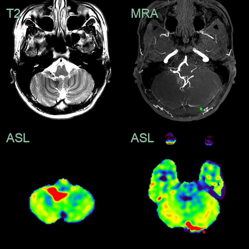
- A 30-year-old patient presented with right sided pulsatile tinnitus.
- Time-of-flight angiography showed a hypertrophied right PICA and a small AVM nidus near the fourth ventricle.
- Confirmed with ASL, the lesion was associated with shunting into the left transverse sinus (high signal on ToF MRA and elevated CBF on ASL).
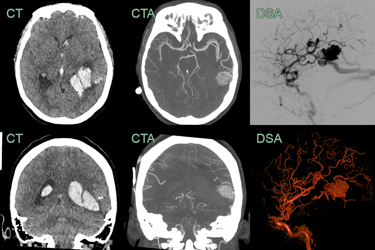
- A 40-year-old patient presented with a headache and right sided weakness.
- CT showed a left temporal haematoma with associated calcification.
- CTA and DSA showed an arteriovenous malformation supplied by the left MCA with cortical drainage.
Treatment
- Options are microsurgical resection, endovascular embolisation and stereotactic radiosurgery, alone or combined
- Choice is guided by Spetzler-Martin grade and rupture status; unruptured low-grade AVMs may be observed (ARUBA)
Differential diagnosis
| Differential Diagnosis | Differentiating Feature |
|---|---|
| Cavernous malformation | Lack of arterial flow on angiography |
| Capillary telangiectasia | Smaller size and less prominent on imaging |
| Developmental venous anomaly | Characteristic "caput medusae" appearance on contrast-enhanced imaging |
| Tumour (e.g., glioma) | Presence of mass effect and surrounding oedema |
| Cerebral aneurysm | Typically appears as a saccular outpouching on a vessel |
| Moyamoya disease | Bilateral involvement of internal carotid arteries with characteristic "puff of smoke" appearance |
| Dural arteriovenous fistula | Direct connection between dural arteries and venous sinuses |
| Sturge-Weber syndrome | Associated facial port-wine stain and leptomeningeal angiomatosis |
| Cerebral abscess | Ring-enhancing lesion with surrounding oedema and fever |
| Multiple sclerosis | Ovoid periventricular white matter lesions on MRI |
-
Spetzler et al. A proposed grading system for arteriovenous malformations. 1986. Journal of neurosurgery - Open in new tab. ↩