Developmental Venous Anomaly (DVA)
Summary
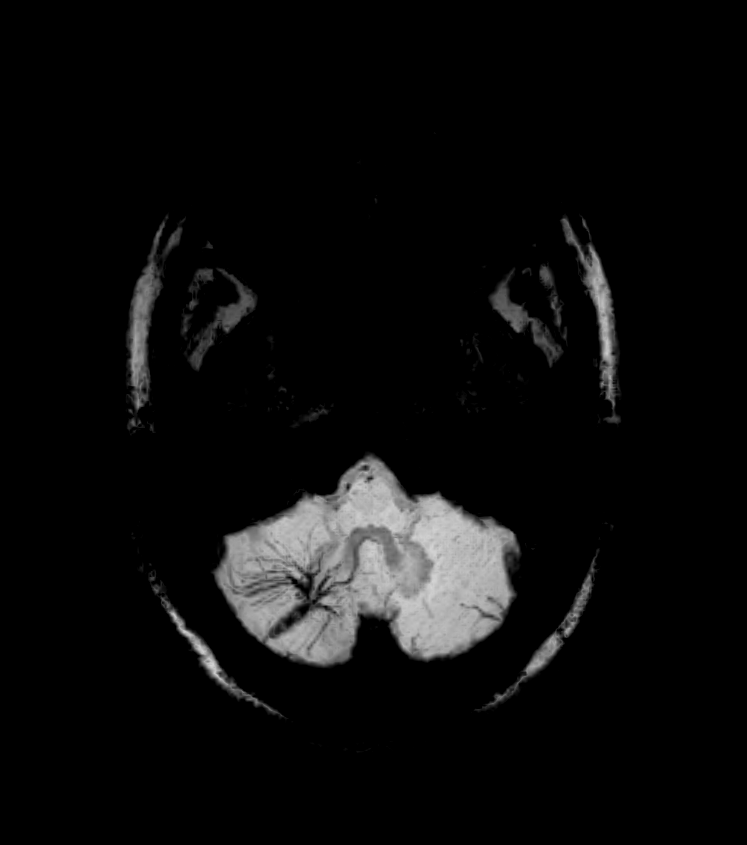
- DVAs are congenital vascular malformations characterised by a radial arrangement of dilated medullary veins converging on a large collecting vein
- Most common cerebral vascular malformation, typically asymptomatic and incidentally discovered on neuroimaging
- Generally considered benign and do not require treatment, but may rarely be associated with other vascular malformations or haemorrhage
Pathophysiology
- Result from arrested development of venous system during embryogenesis
- Represent persistent embryonic medullary veins that failed to regress
- Function as normal drainage pathways for brain parenchyma
- Typically drain into deep or superficial venous systems
Demographics
- Prevalence: 2.6% in autopsy series, up to 6.4% in MRI studies
- No significant gender predilection
- Can occur at any age, but most commonly diagnosed in adults
Diagnosis
- Usually asymptomatic and discovered incidentally on imaging
- Rarely associated with:
- Headaches
- Seizures
- Focal neurological deficits
- May coexist with other vascular malformations (e.g., cavernous malformations)
Imaging
- CT:
- Contrast-enhanced: "caput medusae" appearance of converging veins
- Non-contrast: may show calcifications or associated cavernomas
- MRI:
- T1-weighted: flow void of draining vein
- T2-weighted: hypointense radial veins
- Susceptibility-weighted imaging (SWI): prominent veins
- Post-contrast T1: enhancement of radial veins and draining vein
- Angiography:
- "Caput medusae" appearance in venous phase
- Normal arterial phase
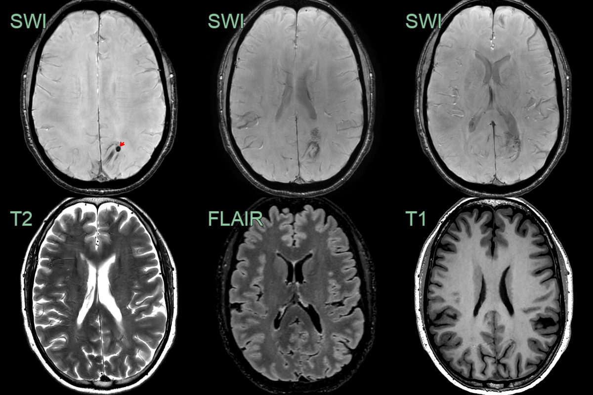
- Incidental findings of a dilated vein with the caput medusa sign.
- The DVA was associated with a small cavernoma (red arrow) .
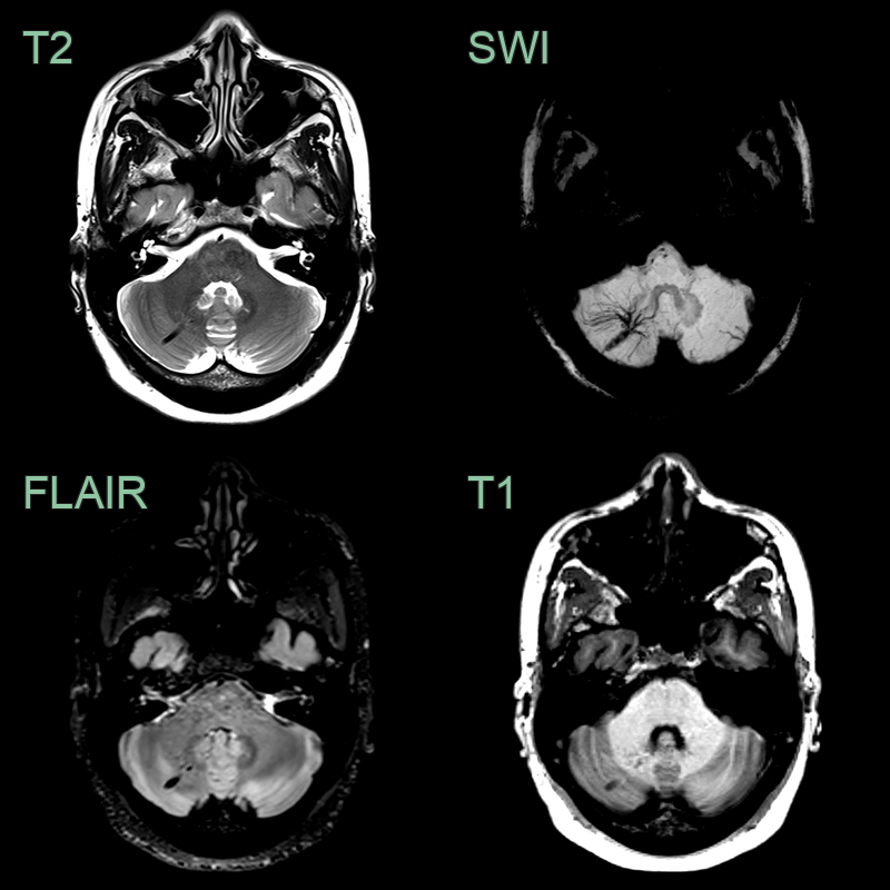
- Incidental finding of a flow void on all sequences with a wider caput medusae on the SWI minimum intensity projection.
Treatment
- Generally, no treatment required for asymptomatic DVAs1
- Management focuses on associated conditions:
- Cavernous malformations: may require surgical resection
- Intracranial haemorrhage: conservative management or surgical intervention based on severity
- Anticoagulation should be used cautiously in patients with DVAs
- Surgical resection of DVAs is contraindicated due to risk of venous infarction
Differential diagnosis
| Imaging differential | Differentiating feature |
|---|---|
| Cavernous malformation | Popcorn lesion with haemosiderin rim; often coexists with a DVA |
| Arteriovenous malformation | Arterial feeders and early venous drainage (shunt) rather than a purely venous phase |
| Capillary telangiectasia | Brush-like SWI signal without a large collecting vein |
| Dural arteriovenous fistula | Arteriovenous shunting with cortical venous reflux |
| Demyelination (central vein) | A single central vein within a lesion, not a radial caput medusae |
-
Hsu et al. Symptomatic Developmental Venous Anomaly: State-of-the-Art Review on Genetics, Pathophysiology, and Imaging Approach to Diagnosis. 2023. AJNR. American journal of neuroradiology - Open in new tab. ↩