Acquired Hepatocerebral Degeneration
Summary
- Neurological syndrome of extrapyramidal and neuropsychiatric symptoms in chronic liver disease
- Occurs with portosystemic shunting and manganese accumulation
- MRI shows symmetric T1 hyperintensity in the globus pallidus without corresponding T2 signal
Pathophysiology
- Manganese deposition in the basal ganglia due to impaired hepatobiliary excretion and portosystemic shunting
- Alzheimer type II astrocytes in the basal ganglia and cortex
- Oxidative stress and mitochondrial dysfunction contribute to neuronal injury
Demographics
- Occurs in a minority of patients with cirrhosis, typically middle-aged to older adults with long-standing disease
- Male predominance
Diagnosis
- Extrapyramidal features (tremor, bradykinesia, rigidity), cognitive/personality change, ataxia and dysarthria
- Deranged liver function; elevated serum ammonia
- Exclude other causes (e.g. Wernicke encephalopathy, Wilson disease)
Imaging
- MRI:
- Symmetric T1 hyperintensity of the globus pallidus, extending to the subthalamic region and substantia nigra
- No corresponding T2/FLAIR signal abnormality
- Signal reflects paramagnetic manganese and usually regresses after transplantation
- CT: typically normal; the T1 change is not attributable to calcification1
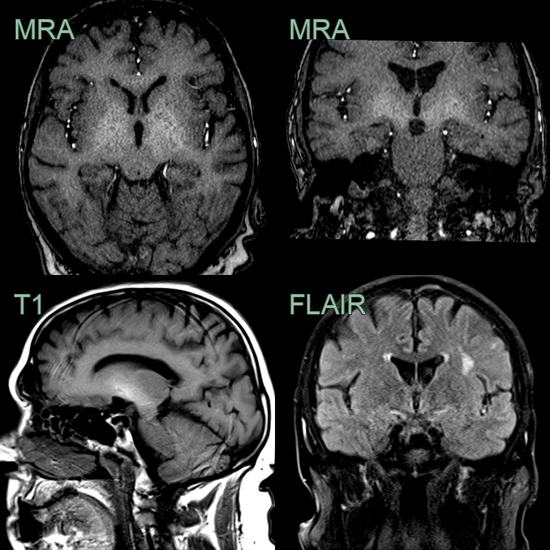
- In a patient with alcoholic liver disease, there is symmetrical T1-hyperintensity in the globi pallidi and subganglionic region.
Treatment
- Treat the underlying liver disease; symptoms and MRI changes may reverse after liver transplantation
- Parkinsonian features respond poorly to levodopa
Differential diagnosis (symmetric T1 hyperintense globus pallidus)
| Imaging differential | Differentiating feature |
|---|---|
| Manganese toxicity (prolonged parenteral nutrition) | Identical symmetric T1 GP hyperintensity; distinguished only by history |
| Wilson's Disease | May show T1 GP signal but also T2 change in putamen/midbrain ("face of the giant panda") |
| Neurofibromatosis type 1 | T1/T2 hyperintense foci (FASI) in the globus pallidus, with other NF1 features |
| Basal ganglia calcification (Fahr) | Dense on CT and blooms on SWI, unlike paramagnetic manganese |
| Kernicterus | Symmetric GP T1 hyperintensity in a neonate |
-
Meissner et al. Acquired hepatocerebral degeneration. 2011. Handbook of clinical neurology - Open in new tab. ↩