Acute Haemorrhage
Summary
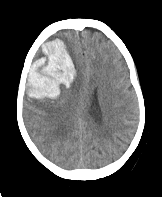
- Acute intracranial haemorrhage: extravasated blood within the brain parenchyma or extra-axial spaces
- Non-contrast CT is first-line; hyperattenuation reflects clot retraction and haemoglobin concentration
- MRI signal evolves predictably with the oxidation state of haemoglobin
Pathophysiology
- Causes include hypertension, trauma, aneurysm rupture, vascular malformation, cerebral amyloid angiopathy, coagulopathy and haemorrhagic transformation of infarction
- Acute clot is dense on CT owing to retraction and high protein/haemoglobin content; severe anaemia or coagulopathy can render fresh blood iso- to hypodense
Imaging
- CT:
- Acute haematoma is hyperdense (~50–70 HU)
- A swirl sign of hypodense, unretracted blood suggests active bleeding
- The CTA spot sign predicts haematoma expansion1
- MRI (signal governed by haemoglobin state):
- Hyperacute (oxyhaemoglobin): T1 iso/hypointense, T2 hyperintense
- Acute (deoxyhaemoglobin): T1 isointense, T2 hypointense
- Blooming on SWI/GRE at all stages
- Distribution suggests aetiology: deep (hypertensive), lobar (amyloid), or along tracts/contrecoup (trauma)
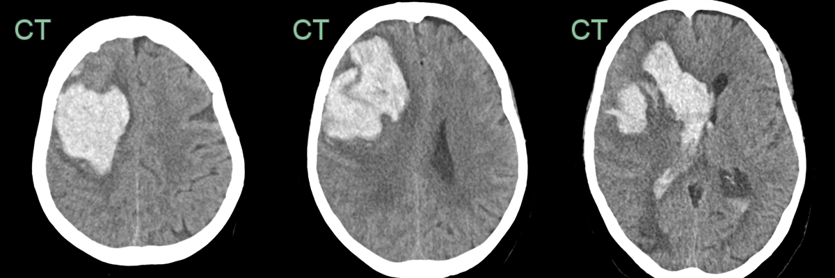
- A hyperdense haematoma, with a thin rim of surrounding oedema, in the right frontal lobe has discharged into the right lateral ventricle.
Treatment
- Blood pressure control and reversal of any anticoagulation
- Surgical evacuation or external ventricular drainage in selected cases
Differential diagnosis
| Differential Diagnosis (and causes of haemorrhage) | Differentiating Feature |
|---|---|
| Haemorrhagic transformation of an infarct | Arterial territory diffusion restriction on MRI beyond the haematoma |
| Subdural Haemorrhage | Crescentic extra-axial collection crossing sutures |
| Epidural Haemorrhage | Biconvex extra-axial collection not crossing sutures |
| Tumour | Mass effect, surrounding oedema, enhancement with contrast |
| Contusion | History of trauma, coup-contrecoup pattern |
| Venous Sinus Thrombosis | Empty delta sign, cord sign on CT/MRI |
| Arteriovenous Malformation | Serpiginous flow voids, calcifications |
| Amyloid Angiopathy | Lobar microhaemorrhages, superficial/lobar distribution |
| Arteriolosclerosis | Basal ganglia small vessel disease and microhaemorrhages |
| Coagulopathy | Abnormal coagulation profile, multiple bleeds |
-
Wada et al. CT angiography "spot sign" predicts hematoma expansion in acute intracerebral hemorrhage. 2007. Stroke - Open in new tab. ↩