Beta Propeller Protein Associated Neurodegeneration
Summary
- A subtype of neurodegeneration with brain iron accumulation (NBIA), caused by WDR45 mutations
- X-linked dominant, usually from de novo mutations, presenting with childhood developmental delay then adult-onset dystonia-parkinsonism and dementia
- The hallmark is T1 hyperintensity of the substantia nigra with a central band of hypointensity ("halo"), plus iron in the substantia nigra and globus pallidus
Pathophysiology
- Caused by mutations in WDR45 gene on X chromosome (Xp11.23)
- WDR45 encodes for WIPI4, a protein involved in autophagy
- Impaired autophagy leads to accumulation of iron and cellular debris
- Neurodegeneration primarily affects basal ganglia and substantia nigra
Demographics
- Rare disorder with estimated prevalence of <1/1,000,000
- Typically presents in childhood or adolescence
- Female predominance due to X-linked dominant inheritance pattern
- Males with germline mutations usually do not survive
Diagnosis
- Clinical features:
- Progressive cognitive decline
- Dystonia
- Parkinsonism
- Seizures
- Sleep disorders
- Genetic testing:
- Identification of pathogenic variants in WDR45 gene
- Biochemical markers:
- Elevated serum ferritin levels
- Normal ceruloplasmin and copper levels
Imaging
- MRI:
- Characteristic T1 hyperintensity of the substantia nigra (± cerebral peduncles) with a central hypointense band1
- Iron-related T2/SWI hypointensity in the substantia nigra and globus pallidus
- Progressive cerebral and cerebellar atrophy
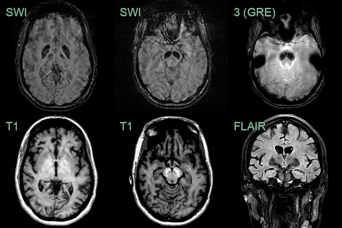
- A 25-year-old patient presented with possible seizures and parkinsonism.
- MRI showed pronounced iron deposition in the globi pallidi and substantia nigra.
- The substantia nigra T1-hyperintensity was typical for the diagnosis of BPAN.
Treatment
- Symptomatic only (levodopa, anticholinergics, anticonvulsants); iron chelation is of unproven benefit
Differential diagnosis
| Differential Diagnosis | Distinguishing Feature |
|---|---|
| Pantothenate Kinase-Associated Neurodegeneration (PKAN) | T2 hypointensity in globus pallidus with central hyperintensity ("eye of the tiger" sign) |
| Mitochondrial Membrane Protein-Associated Neurodegeneration (MPAN) | Linear hypointensity in substantia nigra on T2-weighted images |
| PLA2G6-Associated Neurodegeneration (PLAN) | Cerebellar atrophy and iron accumulation in globus pallidus and substantia nigra |
| Kufor-Rakeb syndrome | Generalized brain atrophy and occasional iron accumulation in basal ganglia |
| Aceruloplasminemia | Widespread iron accumulation in basal ganglia, thalamus, and cerebral cortex |
| Neuroferritinopathy | Cystic degeneration of basal ganglia with iron accumulation |
| Wilson's disease | Putamen/midbrain T2 change ("face of the giant panda"), without the T1 substantia nigra hyperintensity of BPAN |
-
Wilson et al. Consensus clinical management guideline for beta-propeller protein-associated neurodegeneration. 2021. Developmental medicine and child neurology - Open in new tab. ↩