Cholesteatoma
Summary
- Cholesteatoma is a benign but locally destructive lesion of the temporal bone, characterised by an accumulation of keratinizing squamous epithelium
- It typically presents with chronic otorrhea, hearing loss, and potential complications due to erosion of surrounding structures
- Imaging plays a crucial role in diagnosis, surgical planning, and follow-up
Pathophysiology
- Two main types:
- Congenital: Remnant of embryonic epithelial tissue in the middle ear
- Acquired: Most common, develops from retraction pocket in pars flaccida of tympanic membrane
- Growth occurs through accumulation of desquamated keratin and debris
- Expansion leads to bone erosion through pressure necrosis and enzymatic activity
Demographics
- Incidence: 3 per 100,000 in children, 9.2 per 100,000 in adults
- Slightly more common in males
- Peak incidence in second and third decades of life
- Risk factors:
- Chronic otitis media
- Eustachian tube dysfunction
- Craniofacial abnormalities (e.g., cleft palate)
Diagnosis
- Clinical presentation:
- Chronic otorrhea
- Progressive conductive hearing loss
- Otalgia
- Vertigo (in advanced cases)
- Otoscopic examination:
- Retraction pocket or attic perforation
- White or pearly mass behind tympanic membrane
- Audiometry:
- Conductive hearing loss
- Sensorineural component in advanced cases
Imaging
- CT (non-contrast):
- Modality of choice for initial evaluation and surgical planning
- Findings:
- Soft tissue mass in middle ear or mastoid
- Bone erosion (scutum, ossicles, tegmen tympani)
- Widening of aditus ad antrum
- Labyrinthine fistula (in advanced cases)
- MRI:
- Complementary to CT, especially for follow-up and recurrence detection
- Findings:
- T1: Intermediate to low signal intensity
- T2: Heterogeneous, predominantly high signal intensity
- Diffusion-weighted imaging (DWI): Restricted diffusion, high signal on b1000 images
- Non-echo planar diffusion-weighted imaging (non-EPI DWI):
- Higher sensitivity for small cholesteatomas and recurrence detection
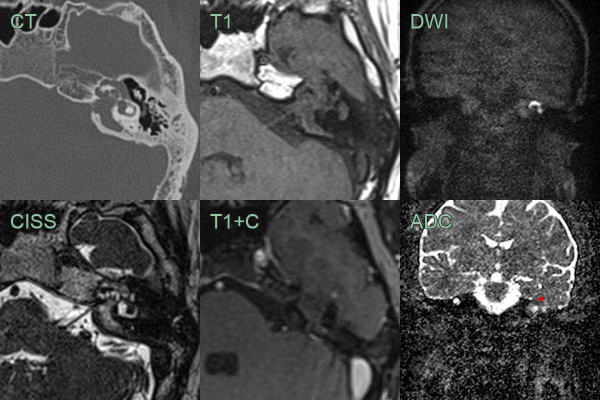
- A 20-year-old patient presented with a facial nerve palsy and sensorineural hearing loss.
- CT showed erosion of the petrous bone including the posterior wall of the internal auditory canal and the wall of the labyrinthine and tympanic facial canal.
- MRI showed a non-enhancing, T2-hyperintense lesion causing diffusion restriction.
Treatment
- Surgical removal (tympanomastoidectomy)1
- Non-EPI DWI is the mainstay for detecting residual/recurrent disease on follow-up, avoiding second-look surgery
- Untreated disease can erode into the labyrinth, facial canal or intracranial compartment
Differential diagnosis
| Differential Diagnosis | Distinguishing Feature |
|---|---|
| Chronic otitis media | Lack of keratin debris on imaging |
| Epidermoid cyst | Markedly restricted diffusion; typically occurs in CPA cistern or other extra-otologic sites |
| Paraganglioma | Characteristic "salt and pepper" appearance on MRI |
| Cholesterol granuloma | Hyperintense on T1-weighted MRI |
| Langerhans cell histiocytosis | Typically multifocal lesions |
-
Yung et al. EAONO/JOS Joint Consensus Statements on the Definitions, Classification and Staging of Middle Ear Cholesteatoma. 2017. The journal of international advanced otology - Open in new tab. ↩