Chordoma
Summary
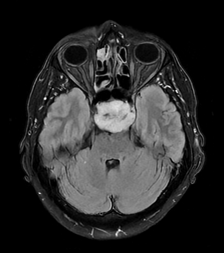
- Rare, slow-growing malignant tumour arising from notochordal remnants
- Typically occurs in the axial skeleton, most commonly at the sacrum and skull base
- Characterised by locally aggressive behaviour and high recurrence rates
Pathophysiology
- Originates from persistent notochordal remnants along the axial skeleton1
- Expresses brachyury, a key transcription factor in notochord development
- Three histological subtypes:
- Conventional (most common)
- Chondroid
- Dedifferentiated (most aggressive)
Demographics
- Incidence: 0.08 per 100,000 person-years
- Median age at diagnosis: 58-60 years
- Slight male predominance (male-to-female ratio 1.5:1)
- Distribution by location:
- Sacrococcygeal: 50-60%
- Skull base: 25-35%
- Mobile spine: 15%
Diagnosis
- Clinical presentation:
- Sacral: pain, neurological deficits, bowel/bladder dysfunction
- Skull base: cranial nerve palsies, headache, visual disturbances
- Histopathology:
- Physaliphorous cells with vacuolated cytoplasm
- Positive immunohistochemistry for brachyury, cytokeratin, and S100 protein
- Genetic testing:
- Duplication of brachyury gene (T) on chromosome 6q27
Imaging
- CT:
- Lytic, destructive lesion with soft tissue mass
- Calcifications in 30-70% of cases
- MRI:
- T1: hypointense to isointense
- T2: hyperintense with heterogeneous signal
- Strong enhancement with gadolinium
- "Honeycomb" appearance due to fibrous septations
- PET/CT:
- Variable FDG uptake, more useful for metastatic disease detection
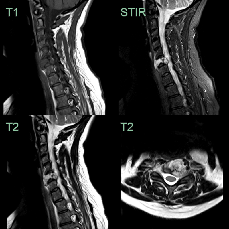
- 50-year-old patient presented with neck pain and a left arm radiculopathy.
- A hyperintense and expansile lesion filling the C6 vertebral body caused compression of the left C7 nerve root.
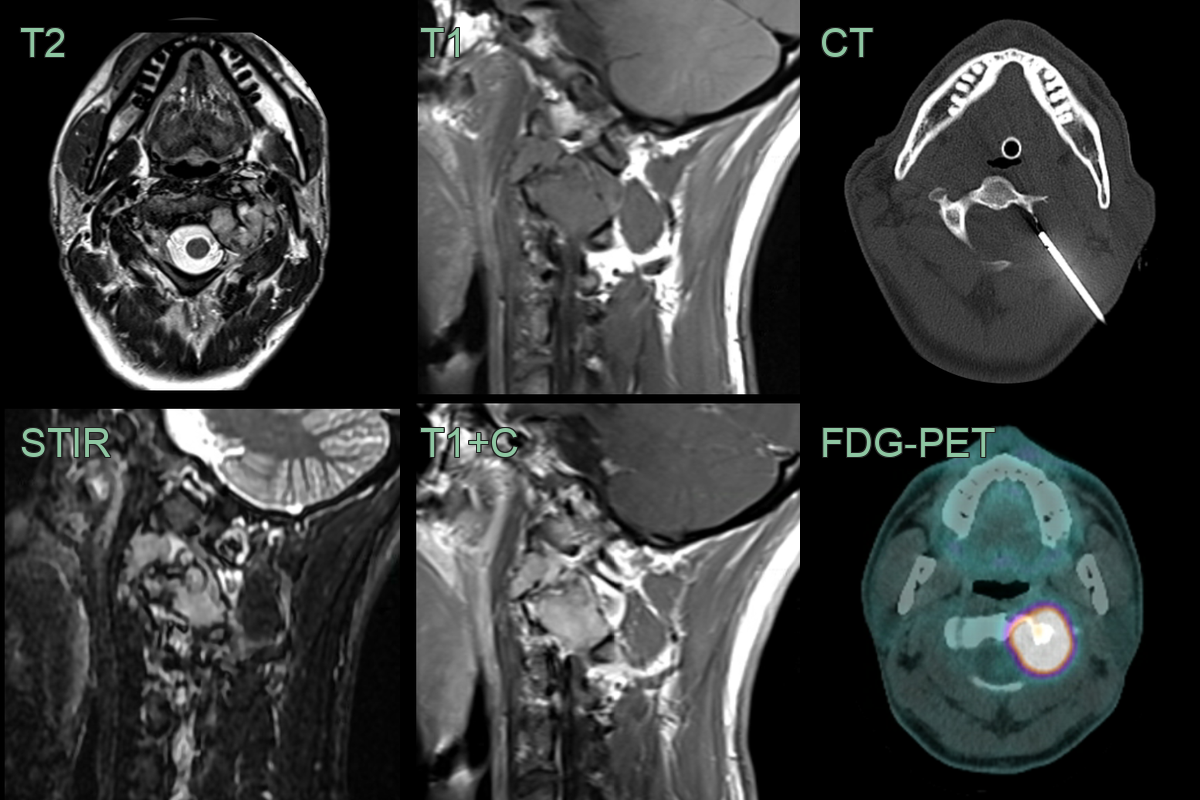
- 30-year-old patient 3 month history of neck pain and crepitus.
- MRI showed an expansile enhancing lesion centred on the left side of the C3 vertebra.
- The lesion was lucent on CT and metabolically active on FDG-PET.
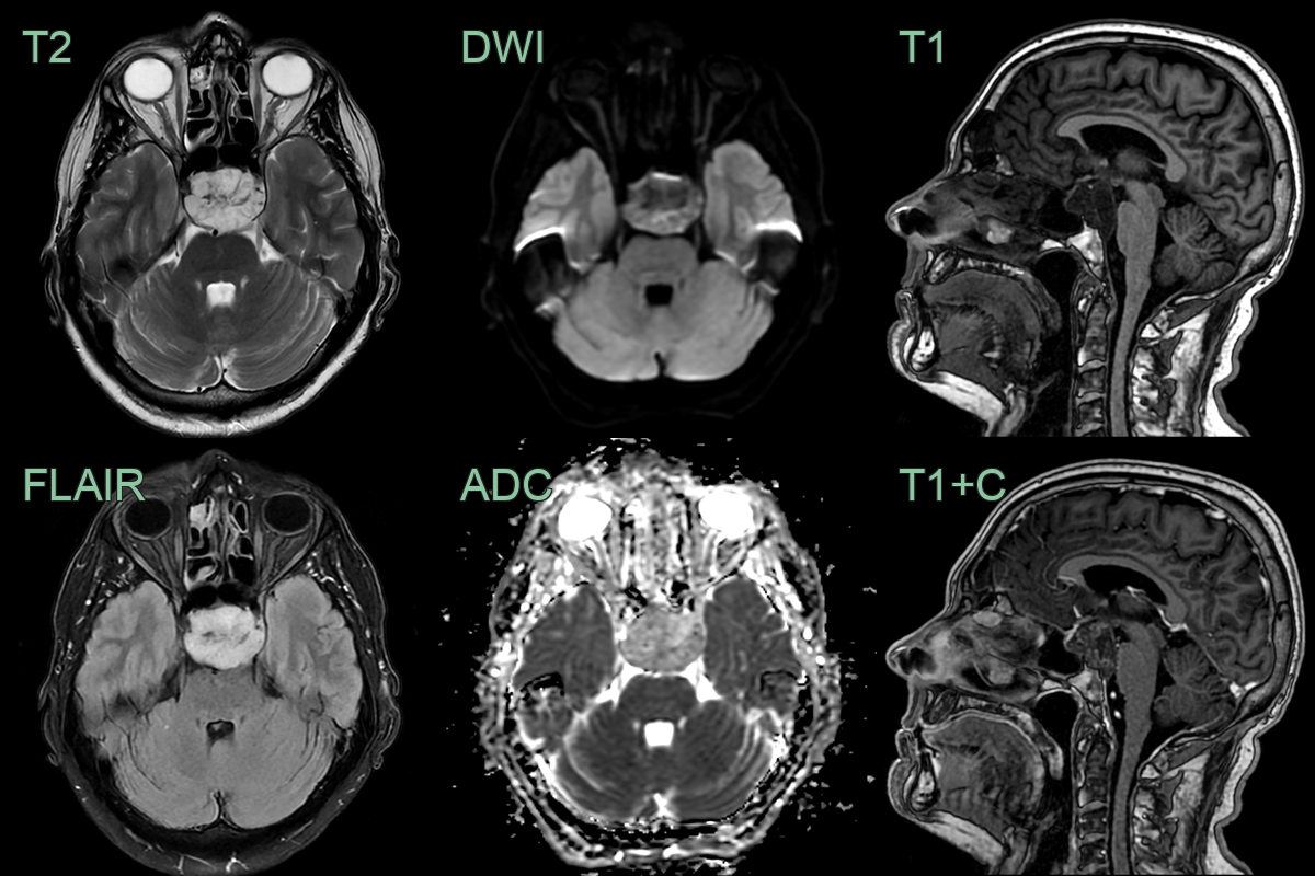
- 60-year-old patient presented with visual impairment and clinical features of hypopituitarism.
- MRI showed a minimally enhancing lesion replacing the superior clivus and pituitary fossa.
- The pituitary gland, infundibular stalk and optic chiasm were compressed.
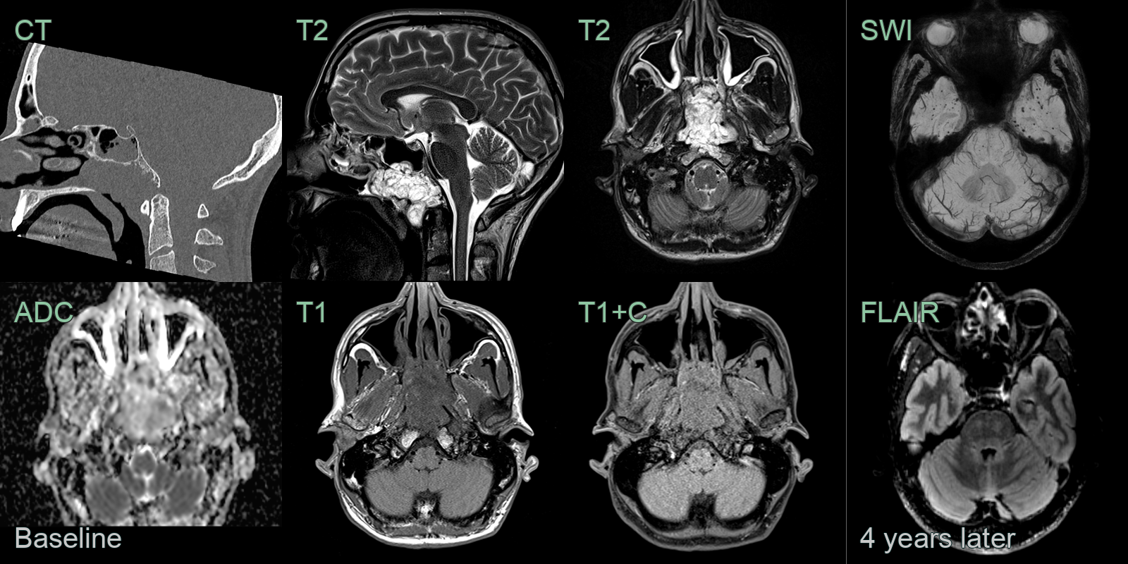
- A 50-year-old patient presented with nasal obstruction.
- MRI showed a lobulated mild enhancing lesion in the nasopharynx with erosion of the inferior cortex of the clivus.
- 4 years later, a follow-up MRI showed no recurrence but many microhaemorrhages in the anterior temporal lobes and brainstem, which were likely to be related to radiotherapy.
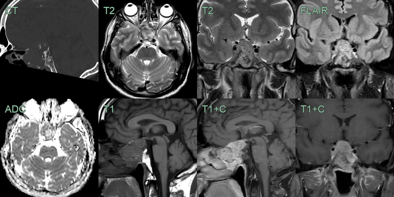
- A 50-year-old patient presented with a visual field defect picked up during a routine eye test.
- CT showed a large destructive lesion centred on the anterior clivus and pituitary fossa.
- MRI showed an avidly enhancing clivus lesion that was compressing the right cisternal optic nerve.
- With the differential including a pituitary macroadenoma, a chordoma was confirmed following a transsphenoidal biopsy.
Treatment
- En bloc resection where feasible, with adjuvant proton beam or carbon ion radiotherapy; high local recurrence rate
Differential diagnosis (clival/midline)
| Imaging differential | Differentiating feature |
|---|---|
| Chondrosarcoma | Off-midline (petro-occipital synchondrosis), with chondroid ring-and-arc calcification |
| Clival metastasis / plasmacytoma | Destructive marrow lesion, often known primary; usually lower T2 signal |
| Pituitary macroadenoma | Centred on the sella and expanding it, rather than destroying the clivus |
| Ecchordosis physaliphora | Small, non-enhancing, retroclival, incidental notochordal rest without bone destruction |
| Skull base meningioma | Dural-based with a dural tail and homogeneous enhancement |
-
Ulici et al. Chordoma. 2022. Archives of pathology & laboratory medicine - Open in new tab. ↩