Chondrosarcoma
Summary
- Malignant cartilaginous tumour arising from bone or soft tissue
- Characterised by production of cartilage matrix
- Imaging shows lobulated, lytic lesion with ring and arc calcifications
Pathophysiology
- Arises from cartilage-forming cells or mesenchymal stem cells
- Classified into three grades based on cellularity, nuclear atypia, and mitotic activity
- Subtypes include conventional, clear cell, dedifferentiated, and mesenchymal chondrosarcoma
Demographics
- Second most common primary bone tumour after osteosarcoma
- Peak incidence in 5th to 7th decades of life
- Slight male predominance (1.5:1)
- Most common sites: pelvis, proximal femur, proximal humerus, and ribs
Diagnosis
- Clinical presentation:
- Pain and swelling at the affected site
- Pathological fracture in advanced cases
- Histopathology:
- Lobulated architecture with hyaline cartilage matrix
- Varying degrees of cellularity and nuclear atypia
-
Molecular markers:
- IDH½ mutations in conventional and dedifferentiated subtypes
-
At the skull base, chondrosarcoma characteristically arises off-midline from the petro-occipital (petroclival) synchondrosis — a key distinction from midline chordoma
Imaging
- CT: lytic, destructive lesion with chondroid ring-and-arc or "popcorn" calcification1
- MRI:
- T1 low-intermediate; very high T2 signal (hyaline cartilage) with a lobulated appearance
- Heterogeneous, often peripheral/septal enhancement
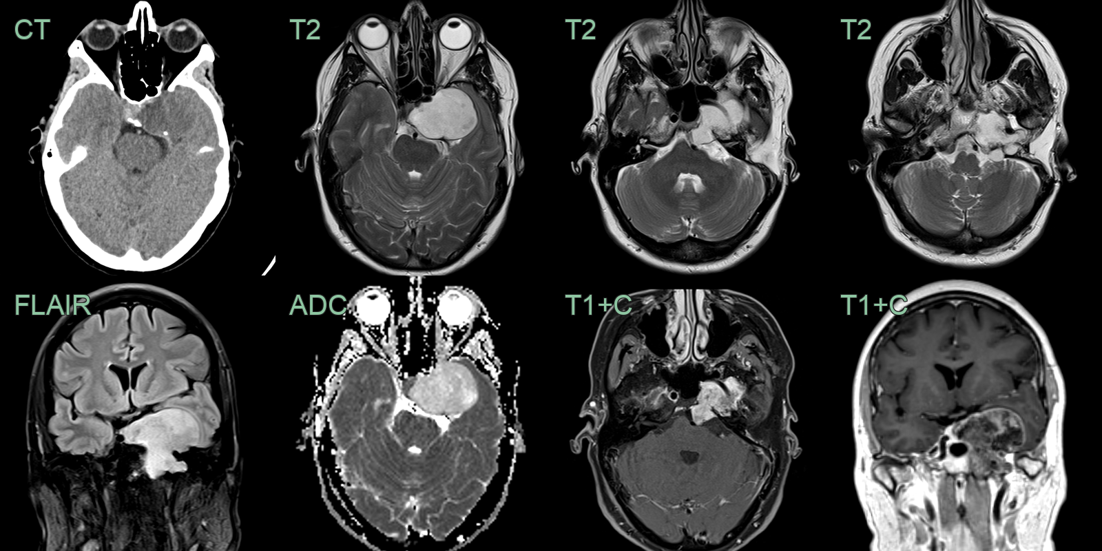
- A 50-year-old patient presented with diplopia.
- Imaging showed a large enhancing non-calcified lesion centred on the left petrous apex with invasion of the cavernous sinus.
Treatment
- Wide surgical resection, usually with adjuvant proton/photon radiotherapy; conventional chemotherapy is of little benefit
Differential diagnosis (skull base)
| Imaging differential | Differentiating feature |
|---|---|
| Chordoma | Midline (clival) rather than off-midline; T2-bright but usually less than chondrosarcoma; brachyury-positive |
| Skull base metastasis / plasmacytoma | Destructive marrow-replacing lesion without chondroid matrix |
| Chondroid meningioma | Dural-based with a dural tail and avid enhancement |
| Fibrous dysplasia | Expansile ground-glass matrix without lytic destruction |
-
Flemming et al. Enchondroma and chondrosarcoma. 2000. Seminars in musculoskeletal radiology - Open in new tab. ↩