Dermoid Cyst
Summary
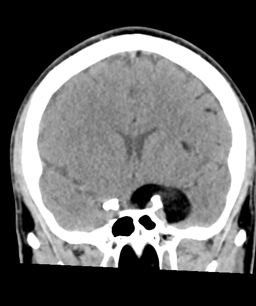
- Benign congenital lesion containing mature tissue derived from ectoderm, mesoderm, and endoderm
- Most commonly found in ovaries, but can occur anywhere along the midline of the body
- Imaging typically shows a cystic mass with fat-fluid levels and calcification
Pathophysiology
- Arise from trapped embryonic germ cells during fetal development
- Contain mature tissues such as:
- Hair follicles
- Sebaceous glands
- Sweat glands
- Teeth
- Bone
- Thyroid tissue
- Lined by keratinized squamous epithelium
- Slow-growing, but can rupture causing inflammation or malignant transformation (rare)
Demographics
- Intracranial dermoids are rare (~0.5% of intracranial tumours), typically midline
- Common sites: parasellar/suprasellar, frontonasal and posterior fossa
Diagnosis
- Often incidental; may present with mass effect or, if ruptured, chemical meningitis or seizures
- Rupture disseminates fat into the subarachnoid space and ventricles
Imaging
- CT: fat-attenuation lesion (−20 to −120 HU), often with mural calcification1
- MRI:
- T1 hyperintense (fat), suppressing on fat-saturated sequences; chemical shift artefact
- Rupture produces scattered T1-hyperintense fat droplets in the sulci and ventricles (fat–fluid levels), a pathognomonic finding
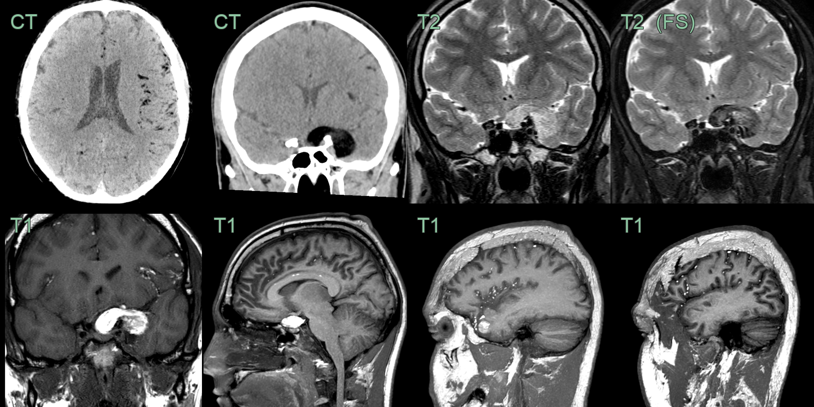
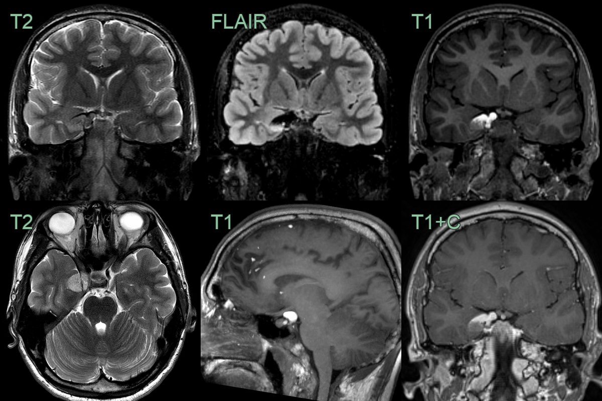
- A 30-year-old patient presented with an acute onset headache.
- MRI showed a lesion in the right cavernous sinus that was T1-hyperintense that suppressed on the fat-suppressed FLAIR imaging, consistent with fat content.
- There were further locules of fat signal over the cerebral hemispheres consistent with dermoid cyst rupture.
Treatment
- Surgical excision with careful avoidance of spillage (which provokes chemical meningitis)
Differential diagnosis
| Imaging differential | Differentiating feature |
|---|---|
| Intracranial lipoma | Homogeneous fat without calcification; does not rupture/disseminate |
| Epidermoid cyst | Follows CSF signal and restricts on DWI; no fat |
| Teratoma | Heterogeneous with fat, calcification and enhancing soft tissue |
| Ruptured Rathke's cleft cyst / craniopharyngioma | T1-bright from proteinaceous content, but no macroscopic fat or disseminated droplets |
-
Pepe et al. Mastoid Dermoid Cyst. 2020. The journal of international advanced otology - Open in new tab. ↩