Free Floating Thrombus
Summary
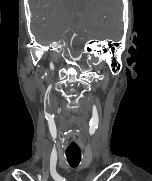
- Free floating thrombus (FFT) is a rare but potentially life-threatening condition characterised by a mobile thrombus in the cardiovascular system, typically attached to a vessel wall by a thin stalk
- FFTs are associated with high risk of embolization and subsequent ischaemic events
- Prompt diagnosis and treatment are crucial to prevent serious complications
Pathophysiology
- An elongated thrombus attached to the vessel wall by a narrow stalk, with mobile intraluminal extension — a source of artery-to-artery embolic stroke
- In the neck it usually arises from a ruptured carotid plaque or a dissection; risk factors include atherosclerosis, hypercoagulable states and malignancy
Demographics
- Incidence is not well-established due to rarity of the condition
- More common in elderly patients
- Higher prevalence in patients with:
- Cardiovascular disease
- History of thromboembolism
- Atrial fibrillation
Diagnosis
- Often an incidental finding on imaging studies
- Clinical presentation may include:
- Symptoms of embolization (e.g., stroke, limb ischaemia)
- Asymptomatic in some cases
- Diagnostic criteria:
- Mobile thrombus
- Thin stalk attachment to vessel wall
- Independent motion from surrounding structures
Imaging
- CTA is the mainstay: an intraluminal filling defect surrounded by contrast (the thrombus is separate from the wall except at its stalk), typically at or just above the carotid bifurcation
- The mobile, non-occlusive nature distinguishes it from fixed plaque; ultrasound may show the thrombus moving with the cardiac cycle
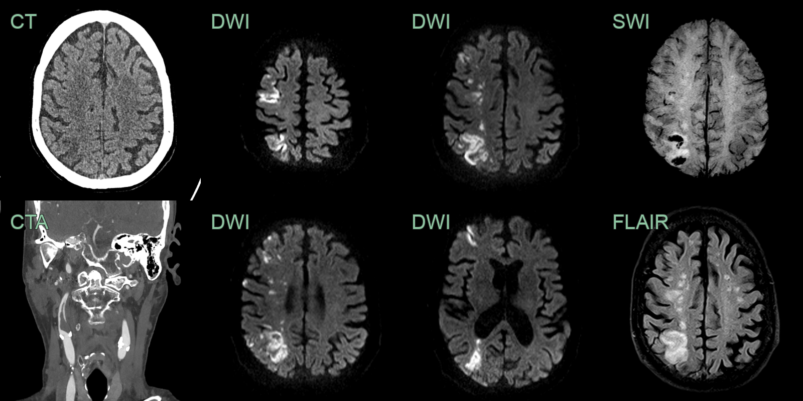
- 60-year-old patient with left sided weakness and inattention.
- CTA showed acute thrombus arising from plaque in the right ICA.
- The filling defect extending distally within the lumen of the vessel was consistent with free floating thrombus.
Treatment
- Anticoagulation is usually first-line; carotid endarterectomy/stenting or thrombectomy for recurrent embolism1
Differential diagnosis
| Imaging differential | Differentiating feature |
|---|---|
| Atherosclerotic plaque | Fixed, calcified, wall-adherent lesion without a mobile intraluminal component |
| Carotid dissection | Crescentic mural haematoma (T1-bright on fat-sat) with a tapered lumen |
| Carotid web | Thin shelf on the posterior bulb wall, not a mobile luminal filling defect |
| Occlusive thrombus | Complete filling defect with no surrounding contrast |
-
Camerotte et al. Efficacy and safety of anticoagulant and antiplatelet therapies in the medical management of carotid free-floating thrombus: A systematic review. 2024. Interventional neuroradiology : journal of peritherapeutic neuroradiology, surgical procedures and related neurosciences - Open in new tab. ↩