Sigmoid Sinus Dehiscence
Summary
- Sigmoid sinus dehiscence is characterised by absence of bone overlying the sigmoid sinus
- Presents with pulsatile tinnitus and may be associated with intracranial hypotension
- Diagnosis relies on high-resolution CT imaging of the temporal bone
Pathophysiology
- Thinning or absence of bone between sigmoid sinus and mastoid air cells
- May be congenital or acquired (e.g. due to increased intracranial pressure)
- Leads to transmission of venous pulsations to surrounding structures
Demographics
- More common in females (female:male ratio approximately 2:1)
- Typically presents in middle-aged adults (40-60 years)
- Prevalence estimated at 1.2% in asymptomatic population
Diagnosis
- Clinical presentation:
- Pulsatile tinnitus (most common symptom)
- Hearing loss
- Vertigo
- Headache
- Otoscopic examination may reveal a bluish mass behind the tympanic membrane1
- Valsalva manoeuvre may alter the intensity of tinnitus
Imaging
- High-resolution CT of the temporal bone:
- Gold standard for diagnosis
- Absence of bone overlying sigmoid sinus
- Thinning of surrounding bone
- Possible protrusion of sinus into mastoid air cells
- MRI:
- T2-weighted images may show flow voids
- MR venography can assess sinus patency and flow
- Digital subtraction angiography:
- Not routinely used but may be helpful in complex cases
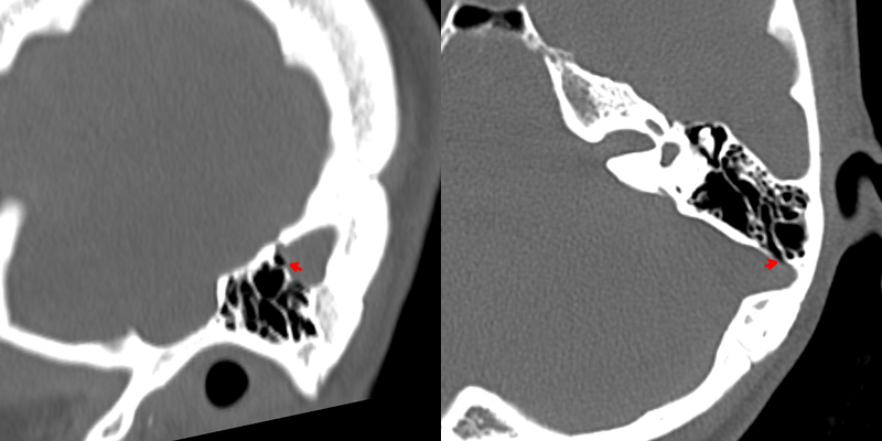
- A 30-year-old female presented with low frequency pulsatile tinnitus that ceased following palpation of the left jugular vein.
- CT showed a focal region of absence of bone over a mastoid air cell, consistent with sigmoid plate dehiscence.
Treatment
- Reassurance, or transmastoid sinus wall resurfacing for disabling pulsatile tinnitus. Look for associated signs of idiopathic intracranial hypertension
Differential diagnosis
| Differential Diagnosis | Distinguishing Feature |
|---|---|
| Glomus tympanicum | Enhancing mass on CT/MRI, "salt and pepper" appearance on T2-weighted MRI |
| Cholesteatoma | Expansile soft tissue mass with bone erosion, non-enhancing on MRI |
| High jugular bulb | Located more inferiorly, smooth margins, no associated symptoms |
| Aberrant internal carotid artery | Anterior location in middle ear on CT; aberrant course lateral to the cochlea |
| Otosclerosis | Lucent halo around cochlea on CT; fissula ante fenestram involvement; no vascular defect |
| Semicircular canal dehiscence | Bony defect involves the superior semicircular canal rather than the sigmoid sinus plate |
| Petrous apex cholesterol granuloma | Expansile lesion in petrous apex, hyperintense on T1-weighted MRI |
| Paraganglioma | Enhancing mass, "salt and pepper" appearance on MRI, may involve jugular foramen |
-
Sotoudeh et al. Prevalence of Sigmoid Sinus Dehiscence and Diverticulum among Adults with Skull Base Cephaloceles. 2020. AJNR. American journal of neuroradiology - Open in new tab. ↩