Superior Semicircular Canal Dehiscence Syndrome
Summary
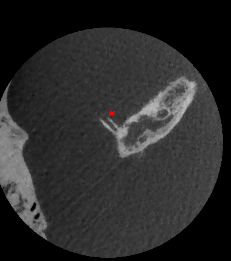
- Rare inner ear disorder characterised by absence of bone overlying the superior semicircular canal
- Presents with vestibular and auditory symptoms triggered by sound or pressure changes
- Diagnosis confirmed by high-resolution CT imaging of temporal bones
Pathophysiology
- Absence of bone over superior semicircular canal creates a "third window" in the inner ear
- Abnormal communication between inner ear and middle cranial fossa
- Results in:
- Pressure-induced displacement of endolymph
- Abnormal activation of vestibular system
- Altered sound transmission to cochlea
Demographics
- Prevalence estimated at 0.5-2% in general population
- Typically presents in middle-aged adults (40-50 years)
- No significant gender predilection
- Bilateral involvement in 25-50% of cases
Diagnosis
- Clinical presentation:
- Vertigo and oscillopsia induced by loud sounds (Tullio phenomenon)1
- Autophony (hearing one's own voice abnormally loud)
- Pulsatile tinnitus
- Chronic disequilibrium
- Diagnostic tests:
- Vestibular evoked myogenic potentials (VEMPs) with lowered thresholds
- Audiometry showing low-frequency air-bone gap
- Fistula test may be positive
Imaging
- High-resolution CT of temporal bones:
- Key diagnostic modality
- Axial and coronal planes with <1mm slice thickness
- Findings:
- Absence of bone over superior semicircular canal
- Direct communication between inner ear and middle cranial fossa
- MRI:
- May show fluid signal extending from superior semicircular canal to middle cranial fossa
- Useful for ruling out other causes of vestibular symptoms
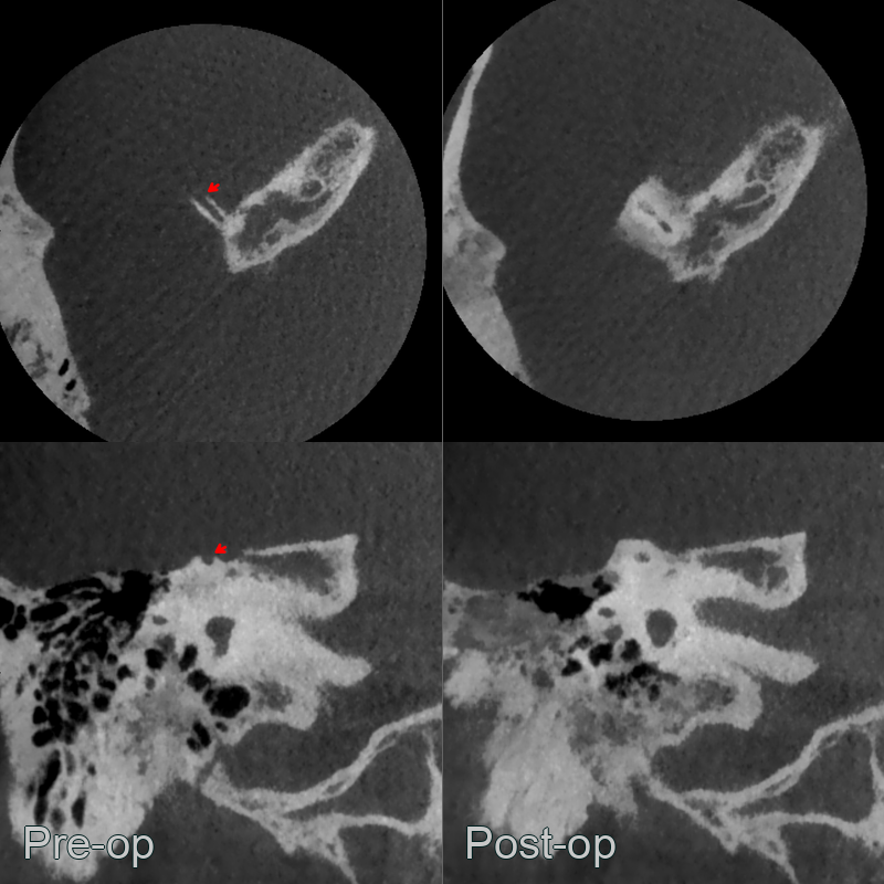
- A 30-year-old patient presented with noise-induced dizziness.
- Cone beam CT showed dehiscence of the right superior semicircular canal.
- The dehiscence resolved following a transmastoid resurfacing with bone pate.
Treatment
- Conservative, or surgical canal plugging/resurfacing for disabling symptoms. Diagnosis rests on thin-section CT reformatted along/perpendicular to the canal to avoid overcalling dehiscence from partial-volume averaging
Differential diagnosis
| Differential Diagnosis | Differentiating Feature |
|---|---|
| Tegmen tympani dehiscence | Bony defect in the tegmen tympani (roof of middle ear) rather than the superior semicircular canal arch on CT; different anatomical location |
| Superior canal thinning (without dehiscence) | Thin but intact bone covering the superior canal on thin-section CT; no true gap; partial volume averaging artefact |
| Enlarged vestibular aqueduct syndrome | Enlarged endolymphatic duct >1.5 mm at midpoint measurement on CT; cochlear involvement |
| Otosclerosis | Lucent halo around cochlea ("halo sign") on CT; fenestral or retrofenestral involvement; no dehiscence of semicircular canal |
| Cholesteatoma | Expansile soft tissue mass with bone erosion in middle ear or mastoid on CT; opacification of middle ear |
-
Steenerson et al. Superior Semicircular Canal Dehiscence Syndrome. 2020. Seminars in neurology - Open in new tab. ↩