Ventriculitis
Summary
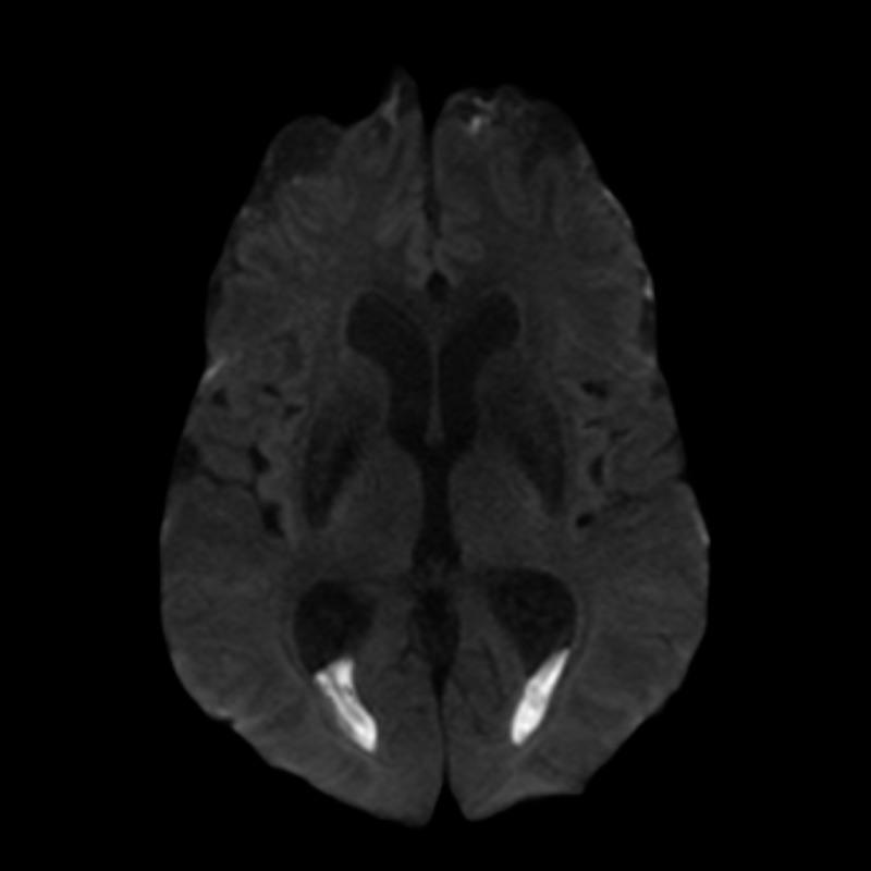
- Inflammation of the cerebral ventricles, often associated with meningitis or intraventricular haemorrhage
- Characterised by ependymal enhancement and intraventricular debris on imaging
- Requires prompt diagnosis and treatment to prevent complications and neurological sequelae
Pathophysiology
- Caused by bacterial, viral, or fungal infections
- Common pathogens:
- Gram-negative bacteria (e.g., E. coli, Klebsiella)
- Staphylococcus species
- Streptococcus species
- Inflammation leads to:
- Ependymal cell damage
- Increased permeability of blood-brain barrier
- Accumulation of inflammatory cells and debris in ventricles
- Can result in hydrocephalus and increased intracranial pressure
Demographics
- Risk factors:
- Neurosurgical procedures (e.g., ventriculostomy, shunt placement)
- Intraventricular haemorrhage
- Prematurity in neonates
- Immunocompromised status
- Incidence:
- 0.8-5.5% following external ventricular drain placement
- Higher in neonates with intraventricular haemorrhage
Diagnosis
- Clinical presentation:
- Fever
- Altered mental status
- Headache
- Neck stiffness
- Seizures
- Laboratory findings:
- Elevated CSF white blood cell count
- Decreased CSF glucose
- Elevated CSF protein
- Microbiological culture of CSF
- Molecular techniques (e.g., PCR) for pathogen identification
Imaging
- CT findings:
- Ventricular dilatation
- Periventricular hypodensity
- Intraventricular debris or air bubbles
- MRI findings:
- T1-weighted images:
- Ependymal enhancement with gadolinium
- Intraventricular debris (hyperintense)
- T2-weighted images:
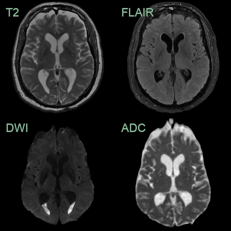
- Periventricular hyperintensity (oedema)
- Intraventricular debris (hypointense)
- FLAIR:
- Hyperintense signal in ventricles
- Periventricular oedema
- Advanced techniques:
- Diffusion-weighted imaging: Restricted diffusion in purulent material
- MR spectroscopy: Elevated lactate and lipid peaks
Treatment
- Antibiotics, CSF drainage and removal of infected devices. Ependymal enhancement with intraventricular debris/fluid levels and restricted diffusion (pus) is the imaging signature1
Differential diagnosis
| Differential Diagnosis | Differentiating Feature |
|---|---|
| Intraventricular haemorrhage | Blood products on CT (hyperdense) and MRI (evolving signal); no ependymal enhancement |
| Leptomeningeal carcinomatosis | Diffuse leptomeningeal and ependymal nodular enhancement; no intraventricular pus or restricted diffusion |
-
Karvouniaris et al. Current Perspectives on the Diagnosis and Management of Healthcare-Associated Ventriculitis and Meningitis. 2022. Infection and drug resistance - Open in new tab. ↩