COL4A1-Related Brain Small Vessel Disease
Summary
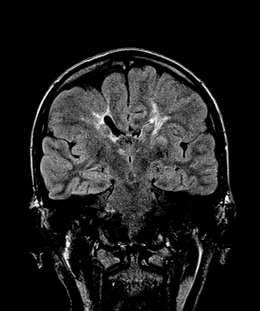
- COL4A1-related brain small vessel disease is a genetic disorder affecting small blood vessels in the brain
- Caused by mutations in the COL4A1 gene, leading to weakened vessel walls and susceptibility to haemorrhage
- Imaging findings include white matter hyperintensities, lacunar infarcts, and microbleeds
Pathophysiology
- COL4A1 gene mutations result in defective type IV collagen production
- Impaired basement membrane integrity in small vessels
- Increased susceptibility to:
- Intracerebral haemorrhage
- Ischaemic stroke
- White matter lesions
Demographics
- Autosomal dominant inheritance pattern
- Variable penetrance and expressivity
- Can affect individuals of all ages, from foetal life to adulthood
- No clear gender predilection reported
Diagnosis
- Clinical presentation:
- Highly variable, ranging from asymptomatic to severe neurological deficits
- Stroke-like episodes
- Migraine with aura
- Seizures
- Cognitive decline
- Genetic testing:
- Sequencing of COL4A1 gene
- Family history assessment
Imaging
- MRI findings:
- White matter hyperintensities on T2-weighted and FLAIR sequences1
- Lacunar infarcts
- Microbleeds on susceptibility-weighted imaging (SWI)
- Enlarged perivascular spaces
- Cerebral microbleeds, particularly in deep and infratentorial regions
- CT findings:
- Hypodensities in white matter
- Evidence of acute haemorrhage in some cases
- Angiography:
- Generally normal appearance of large vessels
- Potential identification of small aneurysms
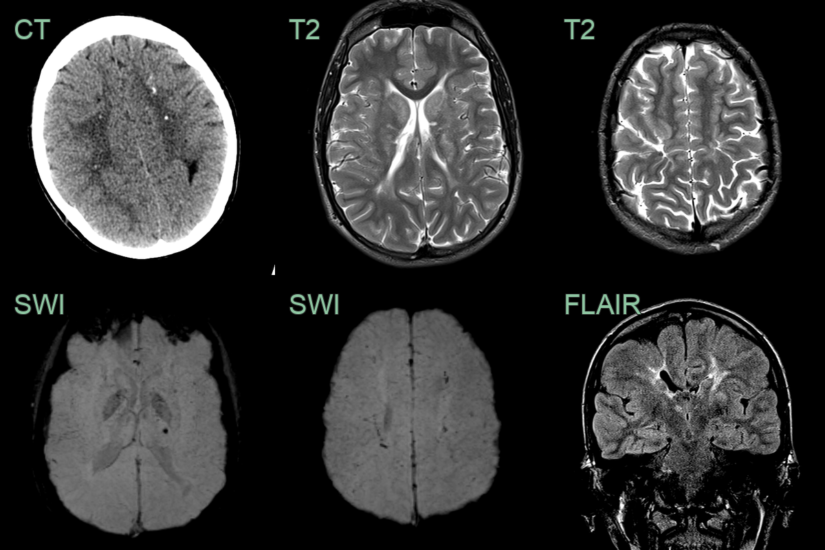
- 20-year-old patient presented with transient sensory disturbance affecting the right arm.
- MRI showed small vessel territory ischaemic damage within the deep grey nuclei and cerebral white matter.
- Cerebral white matter bulk was reduced.
- Abnormal gyration in the perirolandic region suggestive of an intrauterine vascular event.
- Genetic testing revealed a COL4A1 mutation.
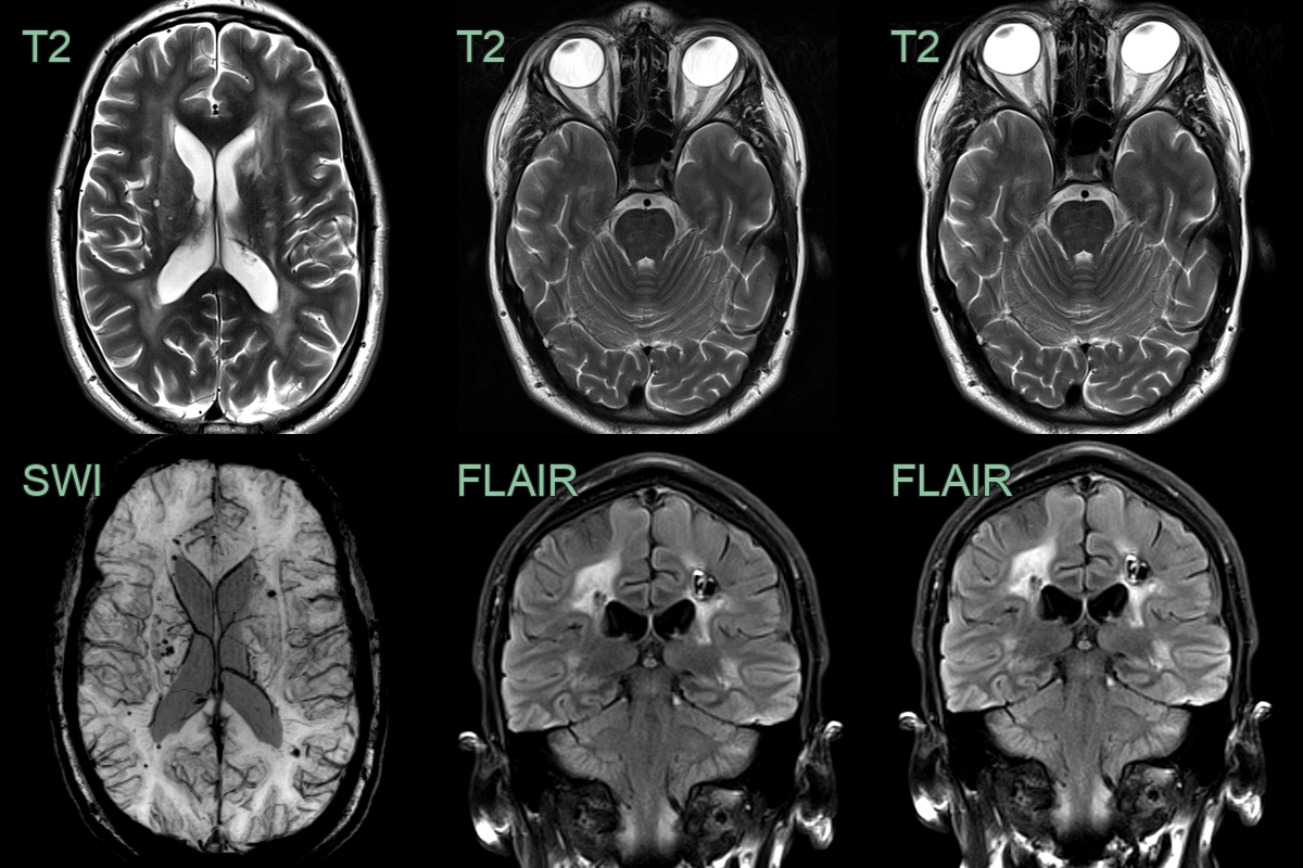
- A 40-year-old patient presented following a left frontoparietal lobar haematoma.
- A follow-up MRI showed a severe burden of small vessel disease, the chronic sequelae of the haematoma and many microhaemorrhages.
- As can be associated with COL4A1, both globes were flattened.
Treatment
- No specific therapy; blood-pressure control and avoidance of anticoagulation given the haemorrhage risk
- Antenatal features (porencephaly, fetal ICH) and associated ocular/renal anomalies are important clues
Differential diagnosis
| Imaging differential | Differentiating feature |
|---|---|
| CADASIL | Anterior temporal pole and external capsule white matter disease |
| Hypertensive microangiopathy | Older patient; lacks the porencephaly/antenatal features of COL4A1 |
| Cerebral amyloid angiopathy | Older; lobar (not deep) microbleeds and superficial siderosis |
| Congenital infection (TORCH) | Parenchymal calcification and malformation; deep microbleeds less typical |
| CARASAL / other hereditary SVD | Overlapping confluent white matter disease; distinguished genetically |
-
Meuwissen et al. The expanding phenotype of COL4A1 and COL4A2 mutations: clinical data on 13 newly identified families and a review of the literature. 2015. Genetics in medicine : official journal of the American College of Medical Genetics - Open in new tab. ↩