Diffuse Hemispheric Glioma
Summary
- Rare, aggressive glioma of adolescents/young adults with diffuse infiltration of a cerebral hemisphere
- Typically presents with seizures, focal neurological deficits, and raised intracranial pressure
- MRI shows unilateral hemispheric involvement with T2/FLAIR hyperintensity and variable enhancement
Pathophysiology
- WHO grade 4 glioma defined by the H3 G34 mutation (H3.3 G34R/V) — distinct from the H3 K27M of diffuse midline glioma
- Arises in the cerebral hemispheres of adolescents/young adults, with diffuse infiltration and poor prognosis
Demographics
- Adolescents and young adults (typically ~15–25 years)
- Rare; recently defined as a distinct WHO 2021 entity
Diagnosis
- Clinical presentation:
- Seizures (focal or generalised)
- Progressive focal neurological deficits
- Signs of raised intracranial pressure (headache, vomiting, papilledema)
- Neuroimaging (MRI) is crucial for initial diagnosis
- Definitive diagnosis requires histopathological examination and molecular testing
Imaging
- MRI is the imaging modality of choice:
- T2/FLAIR: Diffuse hyperintensity involving a large portion of one cerebral hemisphere1
- T1: Hypointense signal in the affected areas
- T1 post-contrast: Variable enhancement patterns, often minimal or absent
- DWI: May show areas of restricted diffusion
- MR spectroscopy: Elevated choline and reduced N-acetylaspartate peaks
- CT:
- May show subtle hypodensity and mass effect
- Calcifications are uncommon
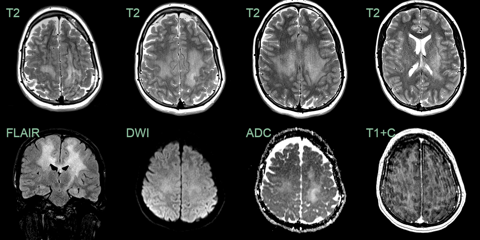
- A 20-year-old patient presented with rapidly progressive spasticity and ataxia.
- MRI showed diffuse bihemispheric ill-defined T2-weighted hyperintensity without diffusion restriction or enhancement.
- Biopsy revealed a H3 G34-mutant diffuse hemispheric glioma.
Treatment
- Maximal safe resection and radiotherapy ± chemotherapy; prognosis remains poor
Differential diagnosis
| Differential Diagnosis | Differentiating Feature |
|---|---|
| Acute disseminated encephalomyelitis (ADEM) | Diffuse bilateral white matter and basal ganglia involvement; no progressive mass effect; no H3K27M imaging correlate |
| Multiple sclerosis | Periventricular ovoid lesions; "Dawson's fingers" on sagittal FLAIR; no hemispheric mass effect |
| Lymphoma | Homogeneous enhancement; periventricular; restricted diffusion; hyperdense on non-contrast CT |
| Metastatic disease | Multiple lesions at grey-white junction; ring or nodular enhancement; surrounding vasogenic oedema |
| Vasculitis | Multifocal cortical and subcortical infarcts in multiple territories; vessel wall enhancement on high-resolution MRI |
| Progressive multifocal leukoencephalopathy (PML) | Subcortical U-fibre involvement; no enhancement; restricted diffusion at active edge; no mass effect |
| Leukodystrophy | Symmetric white matter involvement; specific patterns (anterior, posterior, or central) depending on type |
| Encephalitis | Cortical and limbic T2 signal; often bilateral temporal involvement; may show restricted DWI in active areas |
| Posterior reversible encephalopathy syndrome (PRES) | Posterior-predominant vasogenic oedema; elevated ADC; no hemispheric mass |
| Glioblastoma | Prominent central necrosis with irregular ring enhancement; more established mass effect |
-
Crowell et al. A review of diffuse hemispheric glioma, H3 G34-mutant disease development, DNA repair, microenvironment, and treatments on the horizon. 2025. NPJ precision oncology - Open in new tab. ↩