Multinodular and Vacuolating Neuronal Tumour (MVNT)
Summary
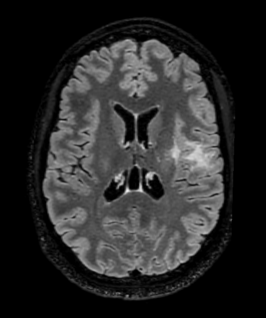
- Rare, benign neuronal tumour characterised by clusters of vacuolated neurons
- Typically located in the cerebral hemispheres, often subcortical
- Usually asymptomatic and incidentally discovered on neuroimaging
Pathophysiology
- Classified as a WHO grade 1 tumour
- Composed of nodules of vacuolated dysplastic neurons
- Immunohistochemistry:
- Positive for HuC/HuD, MAP2, and synaptophysin
- Negative for NeuN, GFAP, and IDH1 R132H mutation
- Molecular profile:
- No specific genetic alterations identified
- MAPK pathway activation suggested
Demographics
- Typically affects adults (mean age 40-50 years)
- No significant gender predilection
- Rare in children
Diagnosis
- Often asymptomatic and incidentally discovered1
- When symptomatic:
- Seizures (most common)
- Headaches
- Cognitive impairment (rare)
- Differential diagnosis:
- Focal cortical dysplasia
- Low-grade glioma
- Dysembryoplastic neuroepithelial tumour (DNET)
Imaging
- MRI characteristics:
- T1: Hypointense to isointense
- T2/FLAIR: Hyperintense, 'bubbly' appearance
- T2*: No susceptibility artefacts
- DWI: No restricted diffusion
- Contrast: Typically non-enhancing
- Location:
- Subcortical white matter
- Commonly in frontal and temporal lobes
- Morphology:
- Multiple small (< 5 mm) nodules
- 'Soap bubble' or 'Swiss cheese' appearance
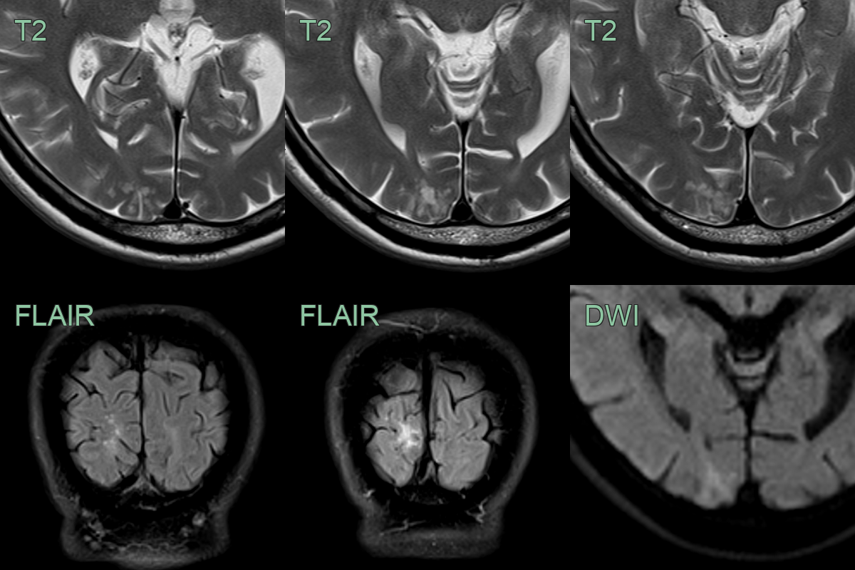
- Incidental T2-hyperintense lesion without mass effect in the right occipital lobe. There was neither diffusion restriction nor pathological enhancement.
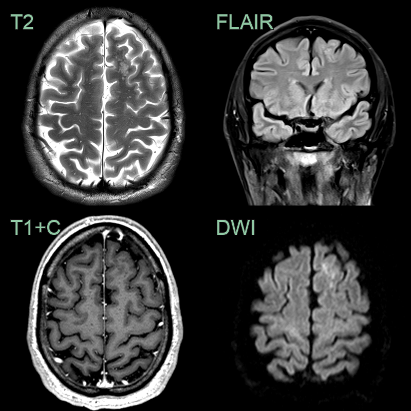
- On an MRI performed for headache, an incidental frontal lobe lesion was identified.
- The bubbly T2-hyperintensity and lack of enhancement are typical of MVNT.
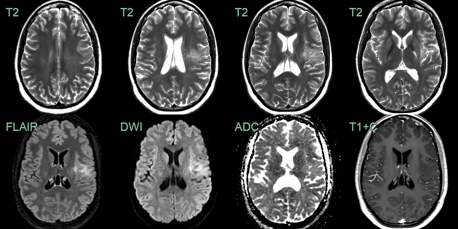
- The "soap bubble" T2-hyperintense lesion in the left cerebral hemisphere without mass effect, diffusion restriction or enhancement was typical of MVNT.
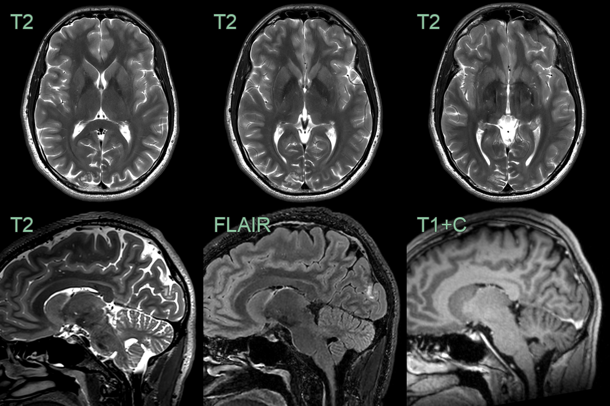
- A 30-year-old patient who present with a headache had an incidental lesion in the right occipital lobe that was consistent with an MVNT.
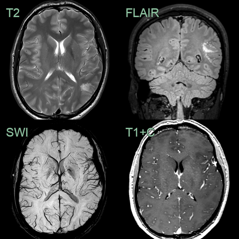
- Clustered subcortical hyperintensities in the right occipital pole were identified incidentally.
- The appearance, lack of enhancement and stability over four years, were consistent with MVNT.
Treatment
- None required; a benign incidental "leave-me-alone" lesion. The bubbly, non-enhancing, FLAIR-hyperintense clusters at the deep cortex/subcortical junction are characteristic
Differential diagnosis
| Differential Diagnosis | Distinguishing Feature |
|---|---|
| Dysembryoplastic neuroepithelial tumour (DNET) | MVNT lacks the specific glioneuronal element seen in DNET |
| Focal cortical dysplasia (FCD) | MVNT has a more nodular appearance and lacks cortical dyslamination typical of FCD |
| Ganglioglioma | MVNT lacks the biphasic pattern of neoplastic glial and neuronal cells seen in ganglioglioma |
| Low-grade glioma | MVNT has a characteristic vacuolated appearance and lacks infiltrative growth |
| Multiple sclerosis plaques | MVNT is typically confined to gray matter, while MS plaques often involve white matter |
| Tuberous sclerosis complex lesions | MVNT lacks calcifications and subependymal nodules typical of TSC |
| Metastatic disease | MVNT is typically non-enhancing and lacks the surrounding oedema often seen with metastases |
| Hamartoma | MVNT has a more organised, nodular structure compared to the disorganised appearance of hamartomas |
| Polymicrogyria | MVNT does not show the characteristic cortical folding abnormalities of polymicrogyria |
| Leukoencephalopathy | MVNT primarily affects gray matter, while leukoencephalopathy involves white matter |
-
Rima et al. Multinodular and vacuolating neuronal tumour: emphasis on expression of early and late neuronal immunomarkers. 2026. Journal of clinical pathology - Open in new tab. ↩