Central Vein Sign
Summary
- Radiological finding in multiple sclerosis (MS) characterised by a central vein within white matter lesions
- Helps differentiate MS from other white matter diseases, particularly small vessel disease
- Visible on susceptibility-weighted imaging (SWI) and T2*-weighted MRI sequences
Imaging Appearance
- Basis of the sign:
- MS lesions develop around small veins in the white matter, with inflammation and demyelination occurring around the central vein
- The vein remains visible within the lesion because of its deoxyhaemoglobin content and the magnetic susceptibility difference between the vein and the surrounding tissue
- Criteria for a positive central vein sign:
- Vein visible in the centre of the lesion
- Vein runs partially or entirely through the lesion
- Vein visible in at least two perpendicular planes
- Sequences:
- Best visualised on susceptibility-weighted imaging (SWI) and T2*-weighted sequences
- FLAIR* imaging (a combination of FLAIR and T2*) can improve detection
- 3T MRI provides better visualisation than 1.5T
- Technique:
- High spatial resolution, thin slices (≤3mm) and minimal slice gap
- Post-processing with minimum intensity projection (mIP); segmented-EPI improves resolution
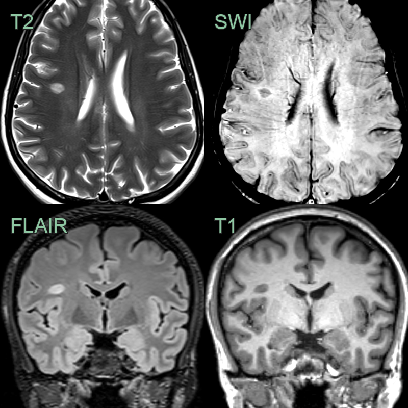
- A patient with a diagnosis of MS had an ovoid lesion in the right frontal subcortical white matter.
- SWI showed that the lesion was centred on a medullary vein.
Clinical Relevance
- A diagnostic aid, not a treatment target: it improves specificity for MS and helps distinguish it from small vessel disease and other white matter disease mimics
- Proposed diagnostic threshold: >40% of lesions showing a central vein supports MS; simplified "select-3/select-6" rules count a fixed number of qualifying lesions1
- Seen in all subtypes of MS — relapsing-remitting, secondary progressive and primary progressive
- Less frequently observed in other white matter diseases
Differential diagnosis
| Differential diagnosis | Differentiating feature |
|---|---|
| Multiple Sclerosis | Central vein sign present in >40% of lesions |
| Small Vessel Disease | Lesions typically do not have central vein |
| Neuromyelitis Optica | Lesions tend to be larger and follow different distribution |
| Acute Disseminated Encephalomyelitis | Lesions are typically larger and more confluent |
| Cerebral Vasculitis | Lesions often follow vascular territories |
| Susac Syndrome | Characteristic involvement of corpus callosum "snowball" lesions |
| Migraine with Aura | No visible lesions on MRI |
| CADASIL | Characteristic involvement of anterior temporal lobes and external capsule |
| Lyme Disease | Lesions are typically non-specific and may resolve with treatment |
| Progressive Multifocal Leukoencephalopathy | Lesions are typically larger and in subcortical white matter |
-
Sati et al. The central vein sign and its clinical evaluation for the diagnosis of multiple sclerosis: a consensus statement from the North American Imaging in Multiple Sclerosis Cooperative. 2016. Nature reviews. Neurology - Open in new tab. ↩